A combined approach to non-carious cervical lesions associated with gingival recession
- Affiliations
-
- 1Department of Conservative Dentistry, Seoul St. Mary's Dental Hospital, College of Medicine, The Catholic University of Korea, Seoul, Korea. dentyeun@catholic.ac.kr
- 2Department of Periodontology, Seoul St. Mary's Dental Hospital, College of Medicine, The Catholic University of Korea, Seoul, Korea.
- KMID: 2346771
- DOI: http://doi.org/10.5395/rde.2016.41.3.218
Abstract
- Non-carious cervical lesions (NCCLs) with gingival recession require specific consideration on both aspects of hard and soft tissue lesion. In the restorative aspect, careful finishing and polishing of the restorations prior to mucogingival surgery is the critical factor contributing to success. Regarding surgery, assessment of the configuration of the lesion and the choice of surgical technique are important factors. The precise diagnosis and the choice of the proper treatment procedure should be made on the basis of both restorative and surgical considerations to ensure the successful treatment of NCCLs.
MeSH Terms
Figure
-
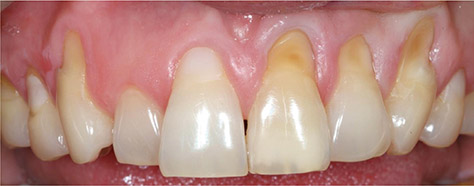
Figure 1 Preoperative frontal view. Note the presence of a non-carious cervical lesion on the maxillary left central incisor, the lateral incisor, and the canines on both sides. Old resin restorations on right first premolar and central incisor showed ill-fitting margins and poor color matching.
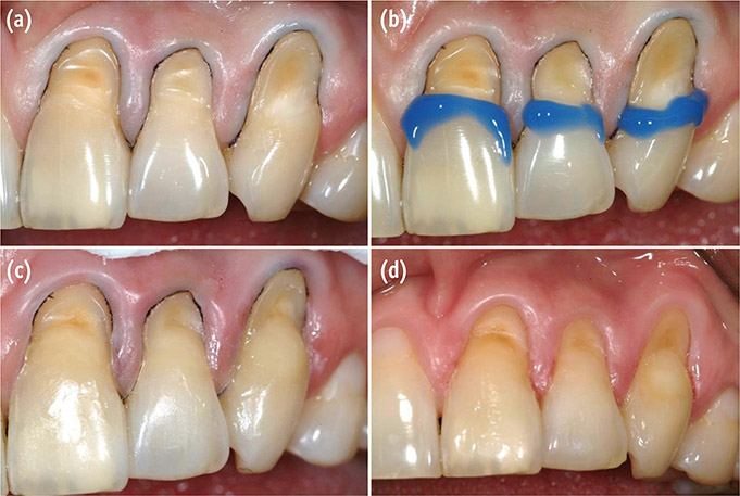
Figure 2 Restorative procedures on the maxillary left side. (a) Field isolation and cavity preparation; (b) Phosphoric acid-selective enamel etching; (c) Adhesive system application and incremental resin filling; (d) Photograph after finishing and polishing.
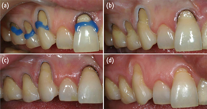
Figure 3 Restorative procedures on the maxillary right side. (a) Phosphoric acid-selective enamel etching after removal of the old restoration and cavity preparation; (b) Adhesive system application; (c) Incremental resin filling; (d) Photograph after finishing and polishing.
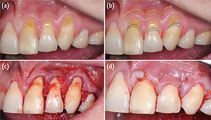
Figure 4 Surgical procedures on the maxillary left side. (a) Multiple adjacent NCCLs; (b) Schematic illustration of the surgery plan; (c) Flap reflection view; (d) Postoperative view after suturing. NCCL, non-carious cervical lesion.
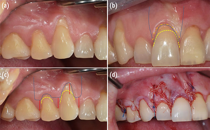
Figure 5 Surgical procedures on the maxillary right side. (a) Multiple adjacent NCCLs; (b and c) Schematic illustration of the surgery plan; (d) Postoperative view after suturing. NCCL, non-carious cervical lesion.
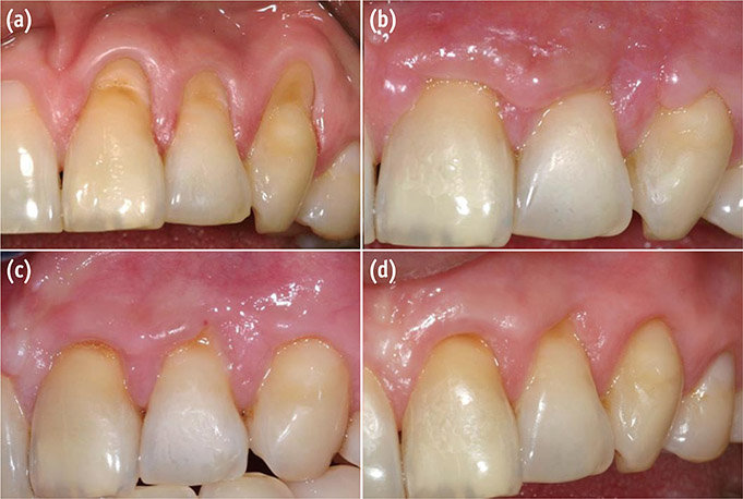
Figure 6 Postoperative view of the maxillary left side. (a) Preoperative view; (b) Four week follow-up; (c) Eight week follow-up; (d) Six month follow-up.
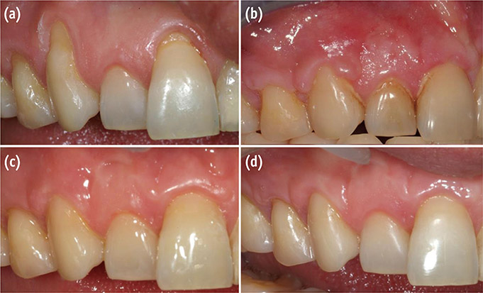
Figure 7 Postoperative view of the maxillary right side. (a) Preoperative view; (b) Four week follow-up; (c) Eight week follow-up; (d) Six month follow-up.

Figure 8 Frontal view. (a) Preoperative view; (b) Postoperative view.
Reference
-
1. Mair LH. Wear in dentistry – current terminology. J Dent. 1992; 20:140–144.2. Aw TC, Lepe X, Johnson GH, Mancl L. Characteristics of noncarious cervical lesions: a clinical investigation. J Am Dent Assoc. 2002; 133:725–733.3. Tyas MJ. Clinical performance of three dentine bonding agents in Class V abrasion lesions without enamel etching. Aust Dent J. 1988; 33:177–180.
Article4. Zucchelli G, Gori G, Mele M, Stefanini M, Mazzotti C, Marzadori M, Montebugnoli L, De Sanctis M. Non-carious cervical lesions associated with gingival recessions: a decision-making process. J Periodontol. 2011; 82:1713–1724.
Article5. Jonck LM, Grobbelaar CJ, Strating H. The biocompatibility of glass-ionomer cement in joint replacement: bulk testing. Clin Mater. 1989; 4:85–107.
Article6. Vandewalle KS, Vigil G. Guidelines for the restoration of Class V lesions. Gen Dent. 1997; 45:254–260.7. Deliberador TM, Martins TM, Furlaneto FA, Klingenfuss M, Bosco AF. Use of connective tissue graft for the coverage of composite resin-restored root surfaces in maxillary central incisors. Quintessence Int. 2012; 43:597–602.8. Lucchesi JA, Santos VR, Amaral CM, Peruzzo DC, Duarte PM. Coronally positioned flap for treatment of restored root surfaces: a 6-month clinical evaluation. J Periodontol. 2007; 78:615–623.
Article9. Santamaria MP, Ambrosano GM, Casati MZ, Nociti FH Jr, Sallum AW, Sallum EA. Connective tissue graft and resin glass ionomer for the treatment of gingival recession associated with noncarious cervical lesions: a case series. Int J Periodontics Restorative Dent. 2011; 31:e57–e63.10. Miller PD Jr. A classification of marginal tissue recession. Int J Periodontics Restorative Dent. 1985; 5:8–13.11. Zucchelli G, Mele M, Stefanini M, Mazzotti C, Mounssif l, Marzadori M, Montebugnoli L. Predetermination of root coverage. J Periodontol. 2010; 81:1019–1026.
Article12. Wennstrom JL. Mucogingival surgery. In : Lang NP, Karring T, editors. Proceedings of the 1st European Workshop on Periodontology. Berlin: Quintessence Publishing Co. Inc.;1994. p. 193–209.13. Camargo PM, Lagos RA, Lekovic V, Wolinsky LE. Soft tissue root coverage as treatment for cervical abrasion and caries. Gen Dent. 2001; 49:299–304.14. Schätzle M, Land NP, Anerud A, Boysen H, Bürgin W, Löe H. The influence of margins of restorations of the periodontal tissues over 26 years. J Clin Periodontol. 2001; 28:57–64.
Article15. Zucchelli G, De Sanctis M. Treatment of multiple recession-type defects in patients with esthetic demands. J Periodontol. 2000; 71:1506–1514.
Article16. Zucchelli G, Testori T, De Sanctis M. Clinical and anatomical factors limiting treatment outcomes of gingival recession: a new method to predetermine the line of root coverage. J Periodontol. 2006; 77:714–721.
Article17. Bignozzi I, Littarru C, Crea A, Vittorini Orgeas G, Landi L. Surgical treatment options for grafting areas of gingival recession association with cervical lesions: a review. J Esthet Restor Dent. 2013; 25:371–382.
Article18. Alkan A, Keskiner I, Yuzbasioglu E. Connective tissue grafting on resin ionomer in localized gingival recession. J Periodontol. 2006; 77:1446–1451.
Article19. Martins TM, Bosco AF, Nóbrega FJ, Nagata MJ, Garcia VG, Fucini SE. Periodontal tissue response to coverage of root cavities restored with resin materials: a histomorphometric study in dogs. J Periodontol. 2007; 78:1075–1082.
Article20. Gomes SC, Miranda LA, Soares I, Oppermann RV. Clinical and histologic evaluation of the periodontal response to restorative procedures in the dog. Int J Periodontics Restorative Dent. 2005; 25:39–47.21. Bruno JF. Connective tissue graft technique assuring wide root coverage. Int J Periodontics Restorative Dent. 1994; 14:126–137.22. Harris RJ. Connective tissue grafts combined with either double pedicle grafts or coronally positioned pedicle grafts: results of 266 consecutively treated defects in 200 patients. Int J Periodontics Restorative Dent. 2002; 22:463–471.23. Langer B, Langer L. Subepithelial connective tissue graft technique for root coverage. J Periodontol. 1985; 56:715–720.
Article
- Full Text Links
-
- Actions
-
Cited
- CITED
-
- Close
- Share
-
- Similar articles
-
- Effective Management of Multiple Non-carious Cervical Lesions with Gingival Recession and Dentin Hypersensitivity: Two Cases Report of Combined Restorative and Periodontal Approach
- Treatment efficacy of gingival recession defects associated with non-carious cervical lesions: a systematic review
- CTG and restoration in treatment of gingival recession associated with a cervical lesion: report of three cases
- Relationship of occlusion and gingival recession
- Correlation between degree of gingival curvature and gingival recession in orthognathic surgery patients
