Primary Cutaneous Adenoid Cystic Carcinoma Arising in Umbilicus
- Affiliations
-
- 1Department of Pathology, Yonsei University College of Medicine, Seoul, Korea. nicekyumi@yuhs.ac
- KMID: 2345559
- DOI: http://doi.org/10.4132/jptm.2015.11.24
Abstract
- No abstract available.
Figure
-
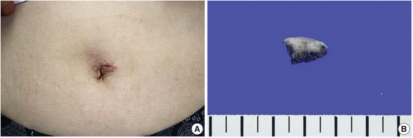
Fig. 1. Macroscopic findings of tumor arising in umbilicus. (A) An ill-defined bean-sized erythematous lesion on the umbilicus. (B) In sections of the specimen from the Mohs micrographic surgery, cut surfaces had an infiltrative firm mass.
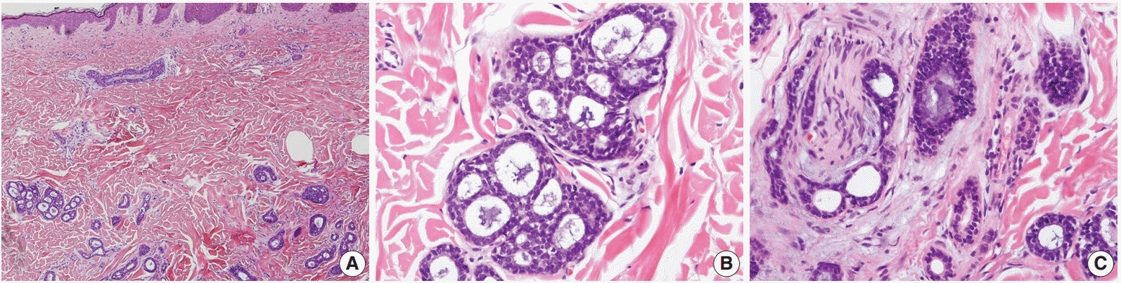
Fig. 2. Microscopic findings of primary cutaneous adenoid cystic carcinoma. (A) Images of basaloid cells that have infiltrated the dermis and the subcutaneous layer, but not the epidermis. Note the tumor cells’ arrangement in nests and their cribriform pattern. (B) Images showing the cribriform and tubular growth patterns of the tumor, which also have a solid portion. (C) Perineural invasion by the tumor.

Fig. 3. Immunohistochemical staining results of primary cutaneous adenoid cystic carcinoma. Immunohistochemical staining images showing strong expression of epithelial membrane antigen (A) and KIT (B) in tumor cells.
Reference
-
1. Boggio R. Letter: adenoid cystic carcinoma of scalp. Arch Dermatol. 1975; 111:793–4.2. Rocas D, Asvesti C, Tsega A, Katafygiotis P, Kanitakis J. Primary adenoid cystic carcinoma of the skin metastatic to the lymph nodes: immunohistochemical study of a new case and literature review. Am J Dermatopathol. 2014; 36:223–8.3. Naylor E, Sarkar P, Perlis CS, Giri D, Gnepp DR, Robinson-Bostom L. Primary cutaneous adenoid cystic carcinoma. J Am Acad Dermatol. 2008; 58:636–41.
Article4. Ramakrishnan R, Chaudhry IH, Ramdial P, et al. Primary cutaneous adenoid cystic carcinoma: a clinicopathologic and immunohistochemical study of 27 cases. Am J Surg Pathol. 2013; 37:1603–11.5. Holst VA, Marshall CE, Moskaluk CA, Frierson HF Jr. KIT protein expression and analysis of c-kit gene mutation in adenoid cystic carcinoma. Mod Pathol. 1999; 12:956–60.6. Jeng YM, Lin CY, Hsu HC. Expression of the c-kit protein is associated with certain subtypes of salivary gland carcinoma. Cancer Lett. 2000; 154:107–11.
Article7. Penner CR, Folpe AL, Budnick SD. C-kit expression distinguishes salivary gland adenoid cystic carcinoma from polymorphous low-grade adenocarcinoma. Mod Pathol. 2002; 15:687–91.
Article8. Mino M, Pilch BZ, Faquin WC. Expression of KIT (CD117) in neoplasms of the head and neck: an ancillary marker for adenoid cystic carcinoma. Mod Pathol. 2003; 16:1224–31.
Article9. Xu YG, Hinshaw M, Longley BJ, Ilyas H, Snow SN. Cutaneous adenoid cystic carcinoma with perineural invasion treated by mohs micrographic surgery-a case report with literature review. J Oncol. 2010; 2010:469049.
Article10. van der Kwast TH, Vuzevski VD, Ramaekers F, Bousema MT, Van Joost T. Primary cutaneous adenoid cystic carcinoma: case report, immunohistochemistry, and review of the literature. Br J Dermatol. 1988; 118:567–77.
Article
- Full Text Links
-
- Actions
-
Cited
- CITED
-
- Close
- Share
-
- Similar articles
-
- Immediate Umbilical Reconstruction after a Mohs Micrographic Surgery for Primary Cutaneous Adenoid Cystic Carcinoma Arising in the Umbilicus
- Skin Metastasis of Adenoid Cystic Carcinoma of Parotid Gland
- A Case of Primary Cutaneous Adenoid Cystic Carcinoma of the Chest Wall
- Primary Cutaneous Adenoid Cystic Carcinoma of the Knee in a Young Male
- Cytopathologic Features of Primary Bronchial Adenoid Cystic Carcinoma: A Case Report
