Vertebra Plana Caused by a Giant Cell Tumor: A Case Report and Literature Review
- Affiliations
-
- 1Department of Radiology, Soonchunhyang University Bucheon Hospital, Korea. mj4907@schbc.ac.kr
- 2Department of Neurosurgery, Soonchunhyang University Bucheon Hospital, Korea.
- 3Department of Pathology, Soonchunhyang University Bucheon Hospital, Korea.
Abstract
- We report here on the case of a 19-year-old woman who presented with progressive weakness of the lower extremities. The radiographs and CT showed vertebra plana of the first thoracic vertebral body. The mass had low signal intensity on the T1-weighted MR image and intermediate signal intensity on the T2-weighted MR image, and this low signal intensity extended to the spinal canal. Histological examination revealed a giant cell tumor (GCT). MR imaging is the imaging modality of choice for helping to distinguish spinal GCT from other spinal tumors by defining the extent and characteristic signal intensity of the tumor.
MeSH Terms
Figure
-
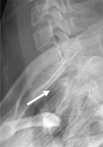
Fig. 1 A 19-year-old woman with spinal GCT. The swimmers lateral view radiograph of the cervical spine, which was acquired with the patient with one arm up and one arm down, shows apparent collapse of the T1 vertebral body.
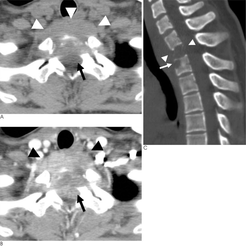
Fig. 2 A 19-year-old woman with a spinal GCT. The precontrast (A) and postcontrast axial CT (B) shows a soft tissue mass (arrowheads) that has destroyed the first thoracic vertebral body with extension to the spinal canal (arrow), and this mass shows homogeneous enhancement (arrowheads) on the contrast-enhanced axial CT (B). The sagittal CT reconstruction (C) shows the paper-thin vertebral body (arrowheads), with preservation of the intervertebral disc space. Round bony erosion is seen in the anterior aspect of the T2 vertebral body (arrow).

Fig. 3 A 19-year-old woman with a spinal GCT. The collapsed first thoracic vertebral body is surrounded by a paravertebral mass with low signal intensity (arrowheads) and the spinal cord is compressed on the T1-weighted image (A), and the mass shows intermediate signal intensity on the T2-weighted image (B) and homogenous enhancement on the contrast-enhanced T1-weighted image (C). MRI shows that the mass involves the anterior aspect of the second thoracic vertebral body (arrows), and this corresponds to the erosive lesion seen on CT (Fig. 2C).
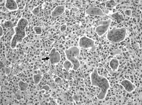
Fig. 4 The histological specimen shows that the tumor consists of mononuclear stromal cells evenly mixed with numerous osteoclast-like giant cells (arrow) (hematoxylin and eosin stain; original magnification, × 200). The mononuclear cells are round to oval or elongated and they lack cytologic atypia. The giant cells are large and they have over 20-30 nuclei.
Reference
-
1. Baghaie M, Gillet P, Dondelinger RF, Flandroy P. Vertebra plana: Benign or malignant lesion? Pediatr Radiol. 1996; 26:431–433.2. Papagelopoulos PJ, Currier BL, Galanis E, Grubb MJ, Pritchard DJ, Ebersold MJ. Vertebra plana caused by primary ewing sarcoma: case report and review of the literature. J Spinal Disord Tech. 2002; 15:252–257.3. Rodallec MH, Feydy A, Larousserie F, Anract P, Campagna R, Babinet A, et al. Diagnostic imaging of solitary tumors of the spine: What to do and say. Radiographics. 2008; 28:1019–1041.4. Murphey MD, Nomikos GC, Flemming DJ, Gannon FH, Temple HT, Kransdorf MJ. From the archives of AFIP. Imaging of giant cell tumor and giant cell reparative granuloma of bone: radiologicpathologic correlation. Radiographics. 2001; 21:1283–1309.5. Hart RA, Boriani S, Biagini R, Currier B, Weinstein JN. A system for surgical staging and management of spine tumors. A clinical outcome study of giant cell tumors of the spine. Spine. 1997; 22:1773–1782.6. Sakurai H, Mitsuhashi N, Hayakawa K, Niibe H. Giant cell tumor of the thoracic spine simulating mediastinal neoplasm. AJNR Am J Neuroradiol. 1999; 20:1723–1726.7. Johnson S, Klostermeier T, Weinstein A. Case report 768. Eosinophilic granuloma of the cervical spine. Skeletal Radiol. 1993; 22:63–65.8. Sanjay BK, Sim FH, Unni KK, McLeod RA, Klassen RA. Giant-cell tumours of the spine. J Bone Joint Surg Br. 1993; 75:148–154.9. Kwon JW, Chung HW, Cho EY, Hong SH, Choi SH, Yoon YC, et al. MRI findings of giant cell tumors of the spine. AJR Am J Roentgenol. 2007; 189:246–250.10. Bidwell JK, Young JW, Khalluff E. Giant cell tumor of the spine: computed tomography appearance and review of the literature. J Comput Tomogr. 1987; 11:307–311.
- Full Text Links
-
- Actions
-
Cited
- CITED
-
- Close
- Share
-
- Similar articles
-
- Giant Cell Tumor Involving the Sixth Cervical Spine: One Case Report
- A Case of the Giant-Cell Tumor in Coccyx
- A Giant Cell Tumor of the Lumbar Vertebra: One Case Report
- Total Spondylectomy for Giant Cell Tumor of Cervical Spine
- Tenosynovial Giant Cell Tumor Showing Severe Bone Erosion in the Finger: Case Report and Review of the Imaging Findings and Their Significance
