J Korean Soc Radiol.
2010 Dec;63(6):531-536.
Serial Changes of the Splenic Volume after Traumatic Intraperitoneal Hemorrhage
- Affiliations
-
- 1Department of Radiology, School of Medicine, Catholic University of Daegu, Korea. yhlee@cu.ac.kr
Abstract
- PURPOSE
We wanted to evaluate the serial changes of the splenic volume in patients with traumatic intraperitoneal hemorrhage.
MATERIALS AND METHODS
20 consecutive patients with traumatic intraperitoneal hemorrhage and who underwent initial CT, early follow-up CT within 30 days and late follow-up CT examinations thereafter were included in this study. The volume of the spleen on each CT examination was measured and the relative splenic volume (RSV) on the initial and early follow-CT examinations was calculated on the basis of the splenic volume on the late follow-up CT. The hemoperitoneum score was calculated on the basis of the size of the intraperitoneal hemorrhage.
RESULTS
The average RSVs of the initial and early follow-up CT were 62.0% and 133.3%, respectively, and all the patients showed an increase of the splenic and relative splenic volumes on the early follow-up CT, as compared with those on the initial CT. Initial splenic contraction was seen in 18 patients (90.0%) and early splenomegaly was seen in 14 patients (70.0%). Patients with initial splenic contraction and early splenomegaly were the most common (12 patient, 60.0%).
CONCLUSION
Initial physiologic splenic contraction was seen in most of the patients with hemoperitoneum, and thereafter early splenomegaly was commonly seen before normalization of the splenic volume.
MeSH Terms
Figure
-
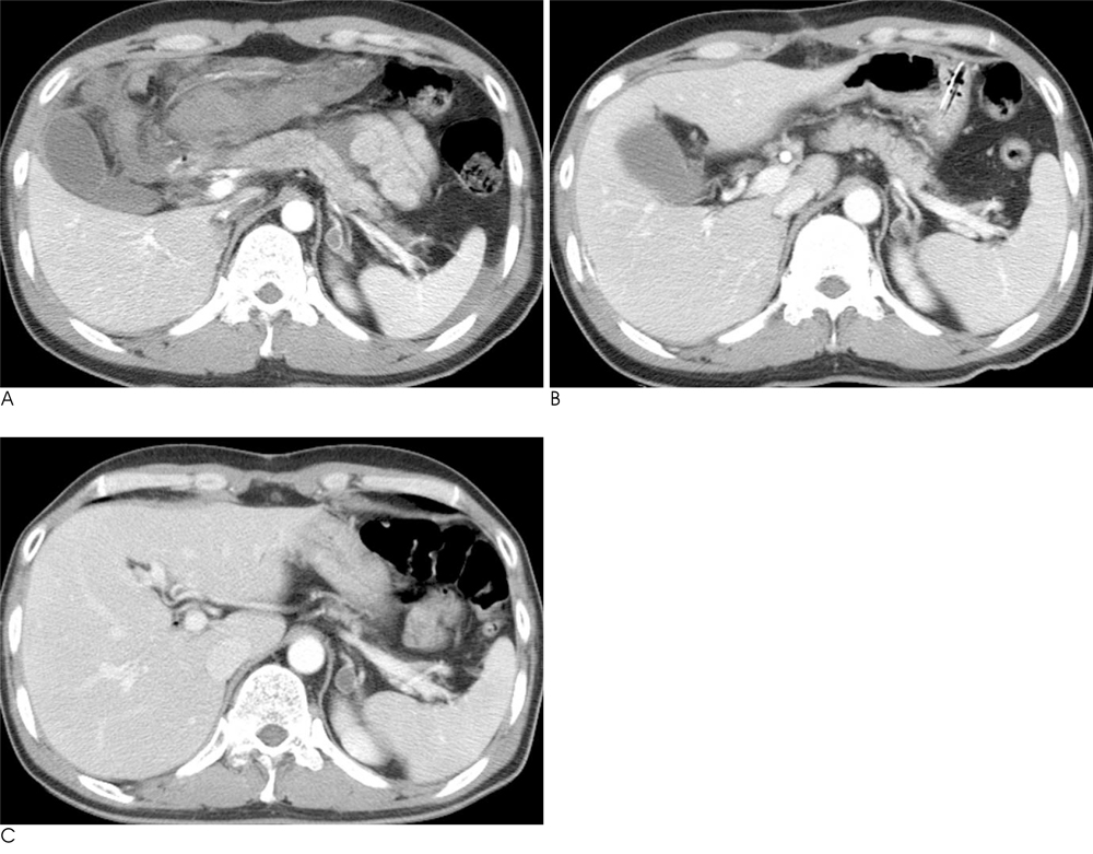
Fig. 1 A 49-year-old man with liver laceration and hemoperitoneum. A. The initial CT showed physiologic splenic contraction (102.7 cc of splenic volume, 54.8% of the relative splenic volume). B. The spleen was enlarged on the follow-up CT taken after 20 days. The splenic volume was 277.9 cc and the relative splenic volume was 148.3%. C. The splenic volume was 187.4 cc on the late follow-up CT after 131 days.
Reference
-
1. Federle MP, Jeffrey RB Jr. Hemoperitoneum studied by computed tomography. Radiology. 1983; 148:187–192.2. Lubner M, Menias C, Rucker C, Bhalla S, Peterson CM, Wang L, et al. Blood in the belly: CT findings of hemoperitoneum. Radiographics. 2007; 27:109–125.3. Shuman WP. CT of blunt abdominal trauma in adults. Radiology. 1997; 205:297–306.4. Goodman LR, Aprahamian C. Changes in splenic size after abdominal trauma. Radiology. 1990; 176:629–632.5. Yaghmai V. Splenic trauma and surgery. In : Gore. RM, Levine MS, editors. Textbook of gastrointestinal radiology. 3rd ed. Philadelphia: Saunders Elsevier;2008. p. 2051–2063.6. Stewart IB, McKenzie DC. The human spleen during physiological stress. Sports Med. 2002; 32:361–369.7. Isbister JP. Physiology and pathophysiology of blood volume regulation. Transfus Sci. 1997; 18:409–423.8. Henderson JM, Heymsfield SB, Horowitz J, Kutner MH. Measurement of liver and spleen volume by computed tomography. Assessment of reproducibility and changes found following a selective distal splenorenal shunt. Radiology. 1981; 141:525–527.9. Staron RB, Ford E. Computed tomographic volumetric calculation reproducibility. Invest Radiol. 1986; 21:272–274.10. Breiman RS, Beck JW, Korobkin M, Glenny R, Akwari OE, Heaston DK, et al. Volume determinations using computed tomography. AJR Am J Roentgenol. 1982; 138:329–333.11. Pozo AL, Godfrey EM, Bowles KM. Splenomegaly: investigation, diagnosis and management. Blood Rev. 2009; 23:105–111.12. Moghimi SM. Mechanisms of splenic clearance of blood cells and particles: towards development of new splenotropic agents. Advanced Drug Delivery Reviews. 1995; 17:103–115.13. Cesta MF. Normal structure, function, and histology of the Spleen. Toxicol Pathol. 2006; 34:455–465.14. Tsushima Y, Tamura T, Tomioka K, Okada C, Kusano S, Endo K. Transient splenomegaly in acute pancreatitis. Br J Radiol. 1999; 72:637–643.
- Full Text Links
-
- Actions
-
Cited
- CITED
-
- Close
- Share
-
- Similar articles
-
- A Case of Spontaneous Hemoperitoneum without Spleen Injury after a Diagnostic Colonoscopy
- Splenic Rupture Complicated by Infective Endocarditis
- Calculation of the Residual Blood Volume after Acute, Non-Ongoing Hemorrhage Using Serial Hematocrit Measurements and the Volume of Isotonic Fluid Infused: Theoretical Hypothesis Generating Study
- Atraumatic Splenic Hemorrhage as a Rare Complication of Pancreatitis: Case Report and Literature Review
- Isolated Traumatic Ganglionic Hemorrhage
