MDCT Findings of Sinus of Valsalva Aneurysms Involving Two Coronary Sinuses: Case Report
- Affiliations
-
- 1Department of Radiology, Inje University Ilsan-Paik Hospital, Korea. sykim@paik.ac.kr
- 2Department of Internal Medicine, Inje University Ilsan-Paik Hospital, Korea.
- 3Department of Cardiothoracic Surgery, Inje University Ilsan-Paik Hospital, Korea.
Abstract
- A sinus of Valsalva aneurysm is relatively rare and usually involves a single sinus. We describe here the multidetector computed tomography features of a case of an unruptured sinus of Valsalva aneurysms that affected the left and noncoronary sinuses in a 51-year-old woman.
MeSH Terms
Figure
-
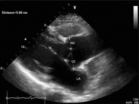
Fig. 1 A transthoracic echocardiography shows the dilatation of the left coronary (LC) and non-coronary (NC) sinuses. The left atrium (LA) is compressed by the dilated left coronary sinus.
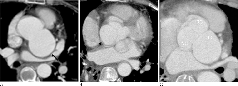
Fig. 2 64 slice-MDCT scans of a 51-year-old woman presenting with chest discomfort for four days. A, B. Two axial images show the unruptured left coronary sinus of Valsalva aneurysm (A, arrow) and the noncoronary sinus of Valsalva aneurysm (B, arrow head). C. The MPR image shows the dilation of the left coronary (LC) and noncoronary (NC) sinus of Valsalva and the normal right coronary sinus (RC).

Fig. 3 A, B. The MPR image (A) and 3-dimensional volume-rendering images (B) show the compression of the left main coronary artery (arrows) by the LC-SVA.
Reference
-
1. Kim KH, Yang TH, Han YC, Cho HJ, Um SJ, Seol SH, et al. Huge aneurysm of the sinus of valsalva compressing the left atrium. J Cardiovasc Ultrasound. 2008; 16:140–142.2. White CS, Plotnick GD. Case 33: sinus of valsalva aneurysm. Radiology. 2001; 219:82–85.3. Thankachen R, Gnanamuthu R, Doshi H, Shukla V, Korula RJ. Unruptured aneurysm of the sinus of valsalva presenting with right ventricular outflow obstruction. Tex Heart Inst J. 2003; 30:152–154.4. Kantarci M, Doganay S, Gundogdu F, Unlu Y. A case with noncoronary sinus of valsalva aneurysm: multidetector computed tomography findings. Heart Surg Forum. 2008; 11:E372–E374.5. Tami LF, Turi ZG, Arbulu A. Sinus of valsalva aneurysms involving both coronary ostia. Cathet Cardiovasc Diagn. 1993; 29:304–308.6. Zannis K, Tzvetkov B, Deux JF, Kirsch EW. Unruptured congenital aneurisms of the right and left sinuses of valsalva. Eur Heart J. 2007; 28:1565.7. Vijayalakshmi IB, Devananda NS, Chitra N. A patient with aneurysms of both aortic coronary sinuses of valsalva obstructing both ventricular outflow tracts. Cardiol Young. 2009; 19:537–539.8. Ho Hwang S, Kim TH, Kim SJ, Kwon HM, Yu KJ. Multidetectorrow computed tomography of a valsalva sinus aneurysm in a patient with behcet disease. J Thorac Imaging. 2006; 21:300–302.9. Matteucci ML, Rescigno G, Capestro F, Torracca L. Syncope triggered by a giant unruptured sinus of valsalva aneurysm. Interact Cardiovasc Thorac Surg. 2009; 9:1047–1048.
- Full Text Links
-
- Actions
-
Cited
- CITED
-
- Close
- Share
-
- Similar articles
-
- Huge Aneurysm of the Sinus of Valsalva Compressing the Left Atrium
- Erratum
- Sudden Death Associated with Anomalous Left Coronary Artery Origin from Right Sinus of Valsalva with Posterior Course
- Two cases of ruptured congenital sinus of Valsalva aneurysms dissecting into the interventricular septum in patients with cerebral infarction
- Transradial Stenting of an Anomalous Right Coronary Artery Originating from the Left Sinus of Valsalva
