Ann Dermatol.
2016 Aug;28(4):511-512. 10.5021/ad.2016.28.4.511.
A Case of Late Stage Sebaceous Trichofolliculoma Showing Overlapping Features with Folliculosebaceous Cystic Hamartoma
- Affiliations
-
- 1Department of Dermatology, College of Medicine, Kyung Hee University, Seoul, Korea. haddal@hanmail.net
- KMID: 2344830
- DOI: http://doi.org/10.5021/ad.2016.28.4.511
Abstract
- No abstract available.
MeSH Terms
Figure
-
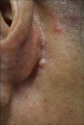
Fig. 1 A solitary asymptomatic 6-mm sized, flesh-colored, dome-shaped nodule on the left postauricular area.
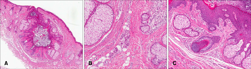
Fig. 2 (A) The epithelial component was surrounded by dense, laminated, collagenous fibroplasia and the fibroepithelial unit was separated by a cleft-like space (H&E, ×40). (B) Mesenchymal stromal components: fibrous tissue with increased blood vessels and mucin deposition, with occasional foci of adipocytes (H&E, ×100). (C) Secondary hair follicles connected to the cystic wall (H&E, ×100).
Reference
-
1. Plewig G. Sebaceous trichofolliculoma. J Cutan Pathol. 1980; 7:394–403.
Article2. Lim HJ, Kim JW, Yu DS. A case of folliculosebaceous cystic hamartoma on the right nasal ala. Ann Dermatol. 2007; 19:170–172.
Article3. Schulz T, Hartschuh W. Folliculo-sebaceous cystic hamartoma is a trichofolliculoma at its very late stage. J Cutan Pathol. 1998; 25:354–364.
Article4. Wu YH. Folliculosebaceous cystic hamartoma or trichofolliculoma? A spectrum of hamartomatous changes inducted by perifollicular stroma in the follicular epithelium. J Cutan Pathol. 2008; 35:843–848.
Article5. Misago N, Kimura T, Toda S, Mori T, Narisawa Y. A revaluation of folliculosebaceous cystic hamartoma: the histopathological and immunohistochemical features. Am J Dermatopathol. 2010; 32:154–161.
Article
