Paravertebral Epithelioid Hemangioendothelioma Associated with Ivory Vertebra: A Case Report
- Affiliations
-
- 1Department of Radiology, Dankook University Hospital, Cheonan, Korea. jyleee@dankook.ac.kr
- 2Department of Neurosurgery, Dankook University Hospital, Cheonan, Korea.
- 3Department of Pathology, Hallym University Sacred Heart Hospital, Anyang, Korea.
- 4Department of Radiology, Dongguk University Gyeongju Hospital, Gyeongju, Korea.
Abstract
- Epithelioid hemangioendotheliomas (EHE) are uncommon and unpredictable vascular tumors that originate in skin, soft tissue, visceral organs, and bones. EHE occurring in the spine has been mostly reported as an osteolytic lesion. To the best of our knowledge, paravertebral EHE with sclerotic bone changes has not been reported in the literature. Here, we report a case of paravertebral EHE accompanied by adjacent sclerotic bone changes in the imaging findings on radiography, CT and magnetic resonance, as well as surgical and pathologic findings.
Figure
-
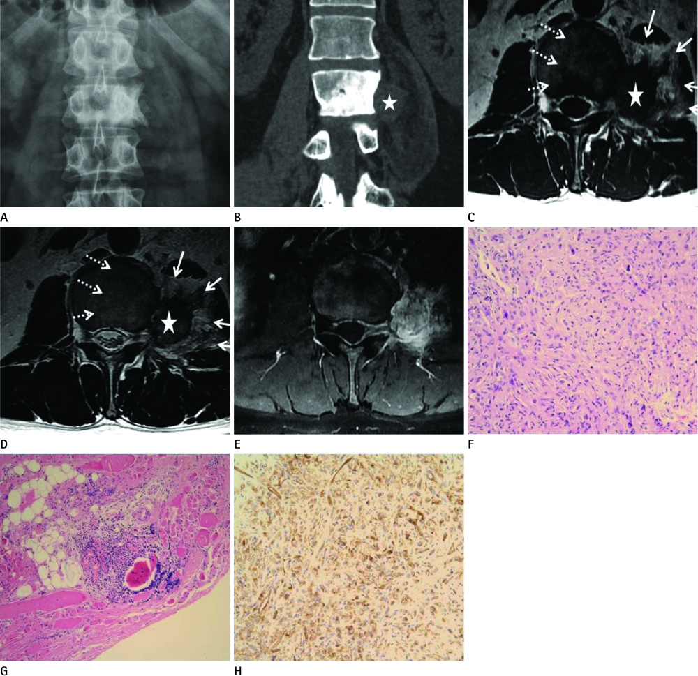
Fig. 1 A 43-year-old man with paravertebral epithelioid hemangioendothelioma. Radiograph with anteroposterior view (A) reveals uniformly increased opacity in the left side of L2 body. Coronal reformatted CT image (B) for the L2 vertebra in the bone window reveals homogeneous increased opacity involving nearly the entire vertebral body. Focal scalloping with sclerotic bony changes in the cortex of L2 is noted in the region abutting the margin of the soft tissue mass (star). MRI with axial T1 weighted image (WI) (C) and T2WI (D) shows a paravertebral solid mass (star) with infiltrative margins and adjacent fat proliferation in the psoas muscle (solid white arrows). A dense radiopaque area on CT shows a mixed signal intensity containing the fat signal (dashed white arrows). The coronal scan with T1WI depicts the relationship of the mass and left L2 spinal nerve (not shown). After gadolinium administration, fat-suppressed T1WI (E) shows intense diffuse enhancement in the mass and mild enhancement in the L2 vertebra and left psoas muscle. Photomicrograph (F) of the specimen shows that the tumor cells are arranged in cord-like pattern within myxohyalinized stroma (Hematoxylin & Eosin, × 200). Photomicrography (G) of the specimen near the tumor shows infiltration of lymphoplasma cells to adipose tissue outside the tumor (× 100). Degenerated muscle fibers that mature adiopocytes and inflammatory cells infiltrate are noted near the tumor. A few muscle fibers are hyalinized and necrotic. Immunohistochemical stain (H) shows that tumor cells were stained with CD31, a representative marker of vascular endothelial cells (× 200).
Reference
-
1. Weiss SW, Enzinger FM. Epithelioid hemangioendothelioma: a vascular tumor often mistaken for a carcinoma. Cancer. 1982; 50:970–981.2. Weiss SW, Goldblum JR. Hemangioendothelioma: vascular tumors of intermediate malignancy. In : Weiss SW, Goldblum JR, editors. Enzinger and Weiss's Soft tissue tumors text book. Philadelphia: MosbyElsvier;2008. p. 681–702.3. Graham TS. The ivory vertebra sign. Radiology. 2005; 235:614–615.4. Gokhan GA, Akyuz M, Gurer IE, Tuncer R. Epithelioid hemangioendothelioma derived from the spine region: case report and review of the literature. Wien Klin Wochenschr. 2006; 118:358–361.5. Christodoulou A, Symeonidis PD, Kapoutsis D, Iordanidis F. Primary epithelioid hemangioendothelioma of the lumbar spine. Spine J. 2008; 8:385–390.6. Abrahams TG, Bula W, Jones M. Epithelioid hemangioendothelioma of bone. A report of two cases and review of the literature. Skeletal Radiol. 1992; 21:509–551.7. Ellis TS, Schwartz A, Starr JK, Riedel CJ. Epithelioid hemangioendothelioma of the lumbar vertebral column: case report and review of literature. Neurosurgery. 1996; 38:402–407.8. Sirikulchayanonta V, Jinawath A, Jaovisidha S. Epithelioid hemangioma involving three contiguous bones: a case report with a review of the literature. Korean J Radiol. 2010; 11:692–696.9. Sung MS, Kang HS, Lee HG. Regional bone changes in deep soft tissue hemangiomas: radiographic and MR features. Skeletal Radiol. 1998; 27:205–210.10. Yao L, Lee JK. Case report 494: hemangioma of surface of ulna with prominent sclerosis. Skeletal Radiol. 1988; 17:378–381.
