Korean Circ J.
2016 Jul;46(4):584-587. 10.4070/kcj.2016.46.4.584.
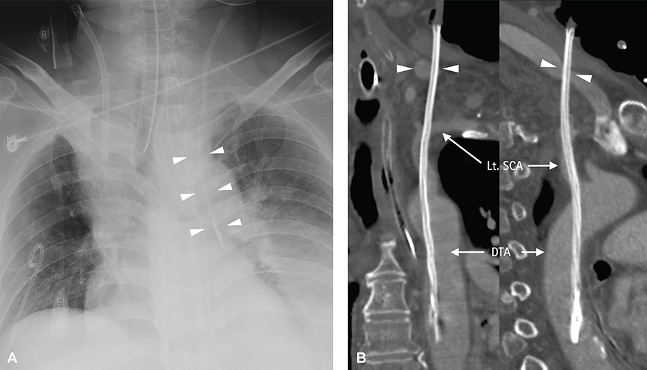
Endovascular Repair Using Suture-Mediated Closure Devices and Balloon Tamponade following Inadvertent Subclavian Artery Catheterization with Large-Caliber Hemodialysis Catheter
- Affiliations
-
- 1Division of Cardiology, Heart Vascular Stroke Institute, Samsung Medical Center, Sungkyunkwan University School of Medicine, Seoul, Korea. cardiochoi@skku.edu
- KMID: 2344437
- DOI: http://doi.org/10.4070/kcj.2016.46.4.584
Abstract
- Accidental subclavian artery cannulation is an uncommon but potentially serious complication of central venous catheterization. Removal of a catheter inadvertently placed in the subclavian artery can lead to substantial bleeding, as achieving hemostasis in this area through manual compression presents considerable difficulty. Additionally, surgical treatment might be unsuitable for high-risk patients due to comorbidities. Here, we report a case of an inadvertently-inserted 11.5-French hemodialysis catheter in the subclavian artery during internal jugular venous catheterization. We performed percutaneous closure of the subclavian artery using three 6-French Perclose Proglide® devices with a balloon tamponade in the proximal part of the subclavian artery. Closure was completed without embolic neurological complications.
MeSH Terms
Figure
Reference
-
1. Rabindranath KS, Kumar E, Shail R, Vaux EC. Ultrasound use for the placement of haemodialysis catheters. Cochrane Database Syst Rev. 2011; CD005279.2. Guilbert MC, Elkouri S, Bracco D, et al. Arterial trauma during central venous catheter insertion: case series, review and proposed algorithm. J Vasc Surg. 2008; 48:918–925. discussion 925.3. Pikwer A, Acosta S, Kölbel T, Malina M, Sonesson B, Akeson J. Management of inadvertent arterial catheterisation associated with central venous access procedures. Eur J Vasc Endovasc Surg. 2009; 38:707–714.4. Eisen LA, Narasimhan M, Berger JS, Mayo PH, Rosen MJ, Schneider RF. Mechanical complications of central venous catheters. J Intensive Care Med. 2006; 21:40–46.5. Schoder M, Cejna M, Hölzenbein T, et al. Elective and emergent endovascular treatment of subclavian artery aneurysms and injuries. J Endovasc Ther. 2003; 10:58–65.6. du Toit DF, Lambrechts AV, Stark H, Warren BL. Long-term results of stent graft treatment of subclavian artery injuries: management of choice for stable patients? J Vasc Surg. 2008; 47:739–743.7. Fraizer MC, Chu WW, Gudjonsson T, Wolff MR. Use of a percutaneous vascular suture device for closure of an inadvertent subclavian artery puncture. Catheter Cardiovasc Interv. 2003; 59:369–371.8. Wallace MJ, Ahrar K. Percutaneous closure of a subclavian artery injury after inadvertent catheterization. J Vasc Interv Radiol. 2001; 12:1227–1230.9. Patel MR, Jneid H, Derdeyn CP, et al. Arteriotomy closure devices for cardiovascular procedures: a scientific statement from the American Heart Association. Circulation. 2010; 122:1882–1893.10. Jahromi BS, Tummala RP, Levy EI. Inadvertent subclavian artery catheter placement complicated by stroke: endovascular management and review. Catheter Cardiovasc Interv. 2009; 73:706–711.11. Berlet MH, Steffen D, Shaughness G, Hanner J. Closure using a surgical closure device of inadvertent subclavian artery punctures during central venous catheter placement. Cardiovasc Intervent Radiol. 2001; 24:122–124.12. Abbott Vascular. Perclose ProGlide Suture-Mediated Closure System. Instructions for use. Unites States: Abbott Vascular;2015. 06. Available from: http://www.abbottvascular.com/ifu.
- Full Text Links
- Actions
-
Cited
- CITED
-
- Close
- Share
- Similar articles
-
- Endovascular Repair of Inadvertent Arterial Injury Induced by Central Venous Catheterization Using a Vascular Closure Device: A Case Report
- Percutaneous Suture-Based Closure Device for Management of Inadvertent Subclavian Artery Catheterization
- Inadvertent Arterial Catheterization of Central Venous Catheter: A Case Report
- Catheter malposition rate following right subclavian venous catheterization
- Unintended Cannulation of the Subclavian Artery in a 65-Year-Old-Female for Temporary Hemodialysis Vascular Access: Management and Prevention


