Korean J Urol.
2015 Oct;56(10):695-702. 10.4111/kju.2015.56.10.695.
Comparison of computed tomography findings between renal oncocytomas and chromophobe renal cell carcinomas
- Affiliations
-
- 1Department of Urology, Urological Science Institute, Yonsei University College of Medicine, Seoul, Korea. urologistyoon@yuhs.ac
- KMID: 2344112
- DOI: http://doi.org/10.4111/kju.2015.56.10.695
Abstract
- PURPOSE
To investigate and distinguish the computed tomography (CT) characteristics of chromophobe renal cell carcinoma (chRCC) and renal oncocytoma.
MATERIALS AND METHODS
Fifty-one patients with renal oncocytoma and 120 patients with chRCC, diagnosed by surgery between November 2005 and June 2015, were studied retrospectively. Two observers, who were urologists and unaware of the pathological results, reviewed the preoperative CT images. The tumors were evaluated for size, laterality, tumor type (ball or bean pattern), central stellate scar, segmental enhancement inversion, and angular interface pattern and tumor complexity. To accurately analyze the mass-enhancing pattern of renal mass, we measured Hounsfield units (HUs) in each phase and analyzed the mean, maximum, and minimum HU values and standard deviations.
RESULTS
There were 51 renal oncocytomas and 120 chRCCs in the study cohort. No differences in clinical and demographic characteristics were observed between the two groups. A central stellate scar and segmental enhancement inversion were more likely in oncocytomas. However, there were no differences in ball-/bean-type categorization, enhancement pattern, and the shape of the interface between the groups. Higher HU values tended to be present in the corticomedullary and nephrogenic phases in oncocytomas than in chRCC. Receiver-operating characteristic curve analysis showed that the presence of a central stellate scar and higher mean HU values in the nephrogenic phase were highly predictive of renal oncocytoma (area under the curve=0.817, p<0.001).
CONCLUSIONS
The appearance of a central stellate scar and higher mean HU values in the nephrogenic phase could be useful to distinguish renal oncocytomas from chRCCs.
MeSH Terms
Figure
-

Fig. 1 Attenuations of 171 renal masses (51 oncocytomas and 120 chromophobe RCCs) on 3-phase computed tomography. HU, Hounsfield unit; RCC, renal cell carcinoma. *p<0.05.
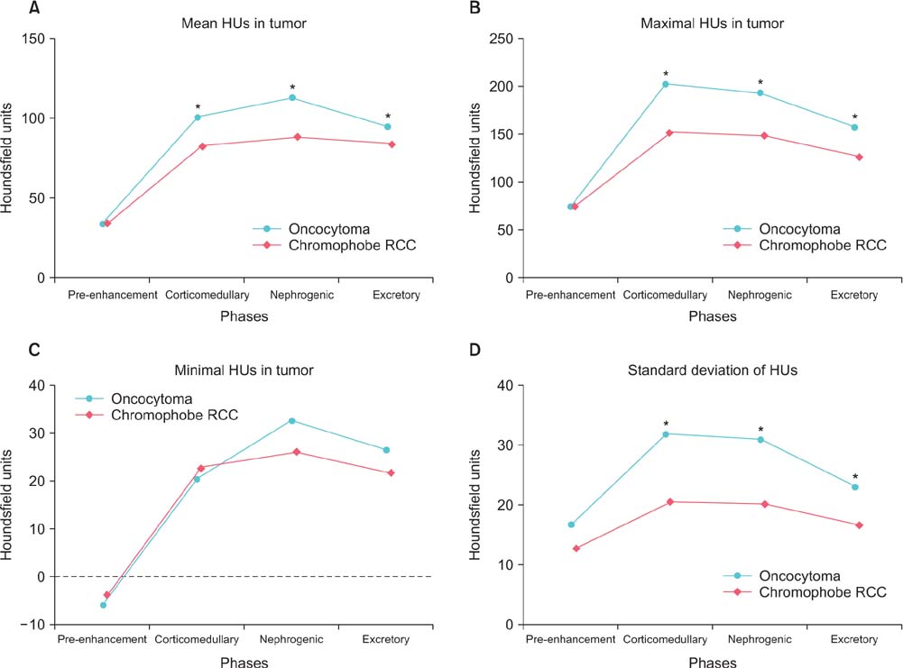
Fig. 2 Attenuations of 84 renal masses (28 oncocytomas and 56 chromophobe RCCs) on 4-phase computed tomography. HU, Hounsfield unit; RCC, renal cell carcinoma. *p<0.05.
Reference
-
1. Cheville JC, Lohse CM, Zincke H, Weaver AL, Blute ML. Comparisons of outcome and prognostic features among histologic subtypes of renal cell carcinoma. Am J Surg Pathol. 2003; 27:612–624.2. Stec R, Grala B, Maczewski M, Bodnar L, Szczylik C. Chromophobe renal cell cancer: review of the literature and potential methods of treating metastatic disease. J Exp Clin Cancer Res. 2009; 28:134.3. Kuroda N, Toi M, Hiroi M, Shuin T, Enzan H. Review of renal oncocytoma with focus on clinical and pathobiological aspects. Histol Histopathol. 2003; 18:935–942.4. Tickoo SK, Amin MB. Discriminant nuclear features of renal oncocytoma and chromophobe renal cell carcinoma. Analysis of their potential utility in the differential diagnosis. Am J Clin Pathol. 1998; 110:782–787.5. Ng KL, Rajandram R, Morais C, Yap NY, Samaratunga H, Gobe GC, et al. Differentiation of oncocytoma from chromophobe renal cell carcinoma (RCC): can novel molecular biomarkers help solve an old problem? J Clin Pathol. 2014; 67:97–104.6. van der Walt JD, Reid HA, Risdon RA, Shaw JH. Renal oncocytoma: a review of the literature and report of an unusual multicentric case. Virchows Arch A Pathol Anat Histopathol. 1983; 398:291–304.7. Muramoto M, Uchida T, Kyuuno H, Ishida H, Utsunomiya T, Egawa S, et al. A case of renal oncocytoma. Hinyokika Kiyo. 1994; 40:47–50.8. Kim JI, Cho JY, Moon KC, Lee HJ, Kim SH. Segmental enhancement inversion at biphasic multidetector CT: characteristic finding of small renal oncocytoma. Radiology. 2009; 252:441–448.9. Woo S, Cho JY, Kim SH, Kim SY. Comparison of segmental enhancement inversion on biphasic MDCT between small renal oncocytomas and chromophobe renal cell carcinomas. AJR Am J Roentgenol. 2013; 201:598–604.10. Dyer R, DiSantis DJ, McClennan BL. Simplified imaging approach for evaluation of the solid renal mass in adults. Radiology. 2008; 247:331–343.11. Kutikov A, Smaldone MC, Egleston BL, Manley BJ, Canter DJ, Simhan J, et al. Anatomic features of enhancing renal masses predict malignant and high-grade pathology: a preoperative nomogram using the RENAL Nephrometry score. Eur Urol. 2011; 60:241–248.12. Mullins JK, Kaouk JH, Bhayani S, Rogers CG, Stifelman MD, Pierorazio PM, et al. Tumor complexity predicts malignant disease for small renal masses. J Urol. 2012; 188:2072–2076.13. Pierorazio PM, Hyams ES, Tsai S, Feng Z, Trock BJ, Mullins JK, et al. Multiphasic enhancement patterns of small renal masses (≤4 cm) on preoperative computed tomography: utility for distinguishing subtypes of renal cell carcinoma, angiomyolipoma, and oncocytoma. Urology. 2013; 81:1265–1271.14. Verma SK, Mitchell DG, Yang R, Roth CG, O'Kane P, Verma M. . Exophytic renal masses: angular interface with renal parenchyma for distinguishing benign from malignant lesions at MR imaging. Radiology. 2010; 255:501–507.15. Kutikov A, Uzzo RG. The R.E.N.A.L. nephrometry score: a comprehensive standardized system for quantitating renal tumor size, location and depth. J Urol. 2009; 182:844–853.16. Lopez-Beltran A, Scarpelli M, Montironi R, Kirkali Z. 2004 WHO classification of the renal tumors of the adults. Eur Urol. 2006; 49:798–805.17. Wu J, Zhu Q, Zhu W, Chen W, Wang S. Comparative study of CT appearances in renal oncocytoma and chromophobe renal cell carcinoma. Acta Radiol. 2015; 05. 13. [Epub]. DOI: 10.1177/0284185115585035.18. Schieda N, McInnes MD, Cao L. Diagnostic accuracy of segmental enhancement inversion for diagnosis of renal oncocytoma at biphasic contrast enhanced CT: systematic review. Eur Radiol. 2014; 24:1421–1429.19. Taura T, Nakamura K, Takashima S, Kaminou T, Yamada R, Shuto T, et al. Heterogeneity of hepatic parenchymal enhancement on computed tomography during arterial portography: quantitative analysis of correlation with severity of hepatic fibrosis. Hepatol Res. 2001; 20:182–192.20. Kojima S, Yoshitomi Y, Yano M, Saotome M, Tanaka K, Endo M, et al. Heterogeneity of renal cortical circulation in hypertension assessed by dynamic computed tomography. Am J Hypertens. 2000; 13(4 Pt 1):346–352.
- Full Text Links
-
- Actions
-
Cited
- CITED
-
- Close
- Share
-
- Similar articles
-
- Differentiation of Chromophobe Renal Cell Carcinoma and Clear Cell Renal Cell Carcinoma by Using Helical CT
- Renal Oncocytoma: Two cases
- Diagnostic Utility of Caveolin-1 and MOC-31 in Distinguishing Chromophobe Renal Cell Carcinoma from Renal Oncocytoma
- Comparison of Ultrasonography, Computed Tomography and Excretory Urography in Staging of Renal Cell Carcinoma
- Expression of E-cadherin in Chromophobe Renal Cell Carcinoma and Its Prognostic Implication
