Intracranial Extraskeletal Myxoid Chondrosarcoma : Case Report and Literature Review
- Affiliations
-
- 1Department of Neurological Surgery, Asan Medical Center, University of Ulsan College of Medicine, Seoul, Korea. jhkim1@amc.seoul.kr
- 2Department of Pathology, Asan Medical Center, University of Ulsan College of Medicine, Seoul, Korea.
Abstract
- Intracranial extraskeletal myxoid chondrosarcoma is extremely rare, with only seven patients previously reported. We present a case report of a 21-year-old woman admitted for weakness in her right extremities and symptoms of increased intracranial pressure. Magnetic resonance imaging (MRI) revealed hydrocephalus and a well-enhanced large mass around her left thalamus. A left parietal craniotomy and a cortisectomy at the superior parietal lobule were performed. Total surgical resection was also performed, and pathology results confirmed an extraskeletal myxoid chondrosarcoma. Postoperative MRI showed no residual tumor, and the patient underwent radiotherapy. After six months of radiotherapy, the patient's headache and weakness had improved to grade IV. This malignant tumor showed high rates of recurrence in previous reports. We here report another occurrence of this highly malignant and rare tumor in a patient treated using total surgical excision and adjuvant radiotherapy.
Keyword
MeSH Terms
Figure
-
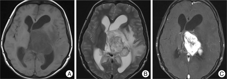
Fig. 1 Imaging findings for the current patient. A : T1-weighted magnetic resonance imaging (MRI) showing homogeneous iso-signal intensity of a 63-mm tumor in the left lateral ventricle along with ventricular dilatation. B : T2-weighted MRI showing a heterogeneous high signal intensity tumor and peritumoral edema. C : T1-weighted enhanced MRI showing strong enhancement of the tumor.
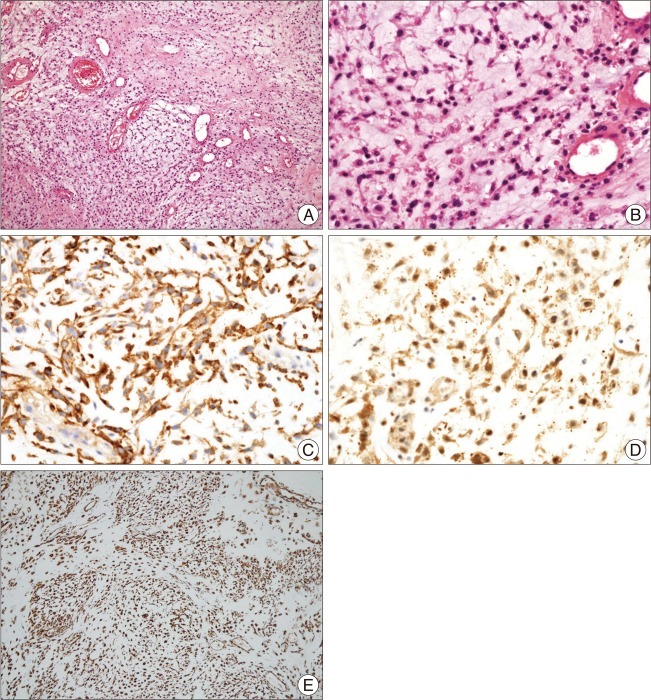
Fig. 2 Histologic features of the tumor in the current patient. A : The tumor consists of strands or cords of oval cells and abundant myxoid stroma (H&E, ×100). B : The tumor cells interconnected to form cords and had relatively uniform oval nuclei and a moderate amount of eosinophilic cytoplasm (H&E, ×400). C, D and E : These tumor cells are positive for epithelial membrane antigen (original magnification ×400) (C), microtubule-associated protein 2 (original magnification ×400) (D), and vimentin (original magnification ×100) (E) by immunohistochemical staining.
Reference
-
1. Bourgouin PM, Tampieri D, Robitaille Y, Robert F, Bergeron D, del Carpio R, et al. Low-Grade Myxoid Chondrosarcoma of the Base of the Skull: CT, MR, and Histopathology. J Comput Assist Tomogr. 1992; 16:268–273. PMID: 1545025.2. Chaskis C, Michotte A, Goossens A, Stadnik T, Koerts G, D'Haens J. Primary intracerebral myxoid chondrosarcoma. Case illustration. J Neurosurg. 2002; 97:228. PMID: 12134922.3. Cummings TJ, Bridge JA, Fukushima T. Extraskeletal myxoid chondrosarcoma of the jugular foramen. Clin Neuropathol. 2004; 23:232–237. PMID: 15581026.4. Enzinger FM, Shiraki M. Extraskeletal myxoid chondrosarcoma. An analysis of 34 cases. Hum Pathol. 1972; 3:421–435. PMID: 4261659.5. González-Lois C, Cuevas C, Abdullah O, Ricoy JR. Intracranial extraskeletal myxoid chondrosarcoma : case report and review of the literature. Acta Neurochir (Wien). 2002; 144:735–740. PMID: 12181708.6. Grossman RI, Davis KR. Cranial computed tomographic appearance of chondrosarcoma of the base of the skull. Radiology. 1981; 141:403–408. PMID: 7291563.
Article7. Hassounah M, Al-Mefty O, Akhtar M, Jinkins JR, Fox JL. Primary cranial and intracranial chondrosarcoma. A survey. Acta Neurochir (Wien). 1985; 78:123–132. PMID: 3911744.8. Im SH, Kim DG, Park IA, Chi JG. Primary intracranial myxoid chondrosarcoma : report of a case and review of the literature. J Korean Med Sci. 2003; 18:301–307. PMID: 12692436.
Article9. Korten AG, ter Berg HJ, Spincemaille GH, van der Laan RT, Van de Wel AM. Intracranial chondrosarcoma : review of the literature and report of 15 cases. J Neurol Neurosurg Psychiatry. 1998; 65:88–92. PMID: 9667567.
Article10. O'Brien J, Thornton J, Cawley D, Farrell M, Keohane K, Kaar G, et al. Extraskeletal myxoid chondrosarcoma of the cerebellopontine angle presenting during pregnancy. Br J Neurosurg. 2008; 22:429–432. PMID: 18568733.11. Oliveira AM, Sebo TJ, McGrory JE, Gaffey TA, Rock MG, Nascimento AG. Extraskeletal myxoid chondrosarcoma : a clinicopathologic, immunohistochemical, and ploidy analysis of 23 cases. Mod Pathol. 2000; 13:900–908. PMID: 10955458.
Article12. Sala F, Talacchi A, Beltramello A, Iuzzolino P, Bricolo A. Intracranial myxoid chondrosarcoma with early intradural growth. J Neurosurg Sci. 1998; 42:159–163. PMID: 10192057.13. Salcman M, Scholtz H, Kristt D, Numaguchi Y. Extraskeletal myxoid chondrosarcoma of the falx. Neurosurgery. 1992; 31:344–348. PMID: 1513440.
Article14. Sato K, Kubota T, Yoshida K, Murata H. Intracranial extraskeletal myxoid chondrosarcoma with special reference to lamellar inclusions in the rough endoplasmic reticulum. Acta Neuropathol. 1993; 86:525–528. PMID: 8310804.
Article15. Scott RM, Dickersin R, Wolpert SM, Twitchell T. Myxochondrosarcoma of the fourth ventricle. Case report. J Neurosurg. 1976; 44:386–389. PMID: 1249620.16. Smith TW, Davidson RI. Primary meningeal myxochondrosarcoma presenting as a cerebellar mass : case report. Neurosurgery. 1981; 8:577–581. PMID: 7266799.
- Full Text Links
-
- Actions
-
Cited
- CITED
-
- Close
- Share
-
- Similar articles
-
- Intracranial Extraskeletal Myxoid Chondrosarcoma in Fourth Ventricle
- A Case of Extraskeletal Myxoid Chondrosarcoma of Pelvic cavity
- A Case Report of Extraskeletal Chondrosarcoma
- Primary Intracranial Myxoid Chondrosarcoma: Report of a Case and Review of the Literature
- Intraspinal Myxoid Chondrosarcoma: Case Report
