Two Cases of Acute Spontaneous Resolution in Macula-Off Rhegmatogenous Retinal Detachment
- Affiliations
-
- 1Department of Ophthalmology, Pusan National University School of Medicine, Busan, Korea. oph97@naver.com
- 2Department of Ophthalmology, Pusan National University Yangsan Hospital, Yangsan, Korea.
- 3Biomedical Research Institute, Pusan National University Yangsan Hospital, Yangsan, Korea.
- KMID: 2339058
- DOI: http://doi.org/10.3341/jkos.2015.56.3.466
Abstract
- PURPOSE
To report 2 cases of acute spontaneous resolution of rhegmatogenous retinal detachment (ASRRRD).
CASE SUMMARY
(Case 1) A 28-year-old male presented with acute visual loss in his left eye for 5 days. The best corrected visual acuity was 10/200 in the left eye and fovea-off retinal detachment with retinal break at the 11-o'clock location was observed. The retina was reattached after 5 days without any treatment. Prophylactic barrier photocoagulation was performed around the break and 3 months after ASRRRD visual acuity improved to 20/30. (Case 2) A 19-year-old male was referred with a history of blurry vision and visual disturbance in his right eye. He underwent a cataract surgery due to traumatic cataract in his right eye 3 years prior. The best corrected visual acuity was 10/200 in the right eye and fovea-off retinal detachment with retinal break at the 10:30-o'clock location was observed. The retina reattached spontaneously after 5 days. Prophylactic barrier photocoagulation was performed around the break and 3 months after ASRRRD visual acuity improved to 20/30.
CONCLUSIONS
It would be better to check the status of fundus and the visual acuity before the surgery in the cases of rhegmatogenous retinal detachment in young age.
MeSH Terms
Figure
-
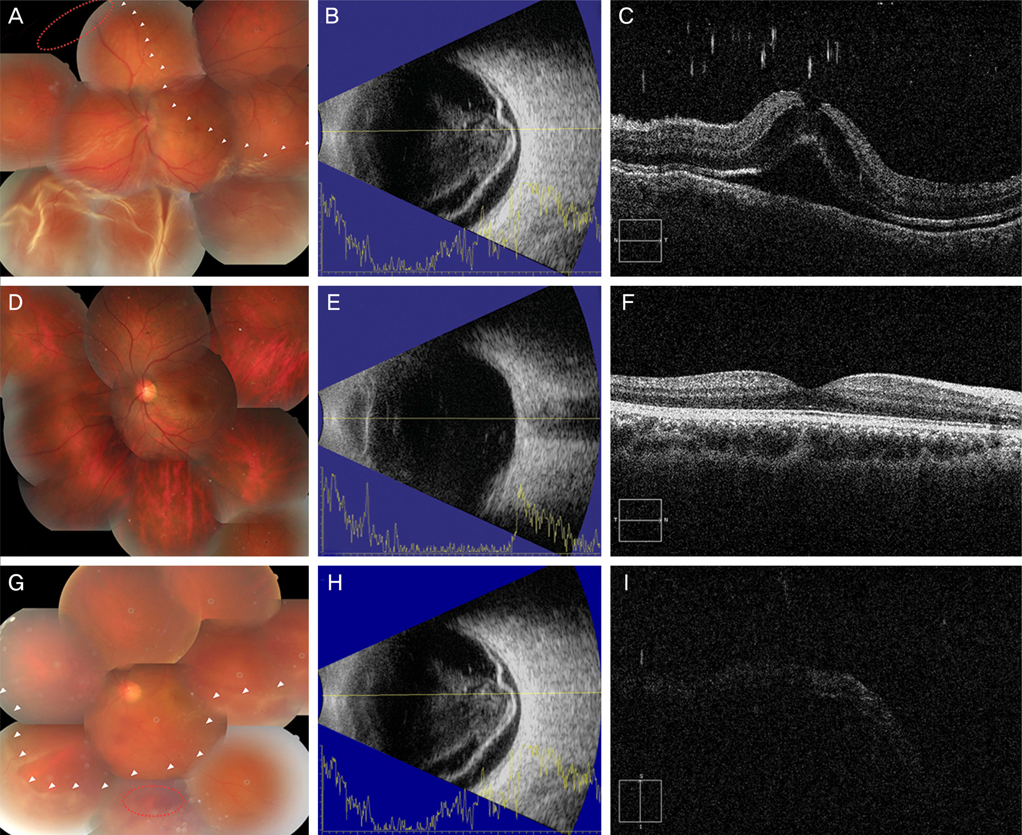
Figure 1. Case 1 patient. (A) Bullous rhegmatogenous retinal detachment is observed at the area inferior to the margin of retinal detachment (white arrow heads) and retinal break is located at 11 o’clock direction anterior to the equator (red dotted circle). (B, C) Macula-off retinal detachment is demonstrated by ultrasonograph and ocular computed tomography (OCT) images. (D) Acute spontaneous reattachment of rhegmatogenous retinal detachment (ASRRRD) after 5 days without any treatment. (E, F) ASRRRD and no definite posterior vitreous detachement are observed in ultrasonograph and OCT images. (G, H, I) Recurrence of rhegmatogenous retinal detachment with dispersive vitreous hemorrhage after blunt trauma was occurred and demonstrated by fundus photograph, ultrasonograph and OCT images. Margin of retinal detachment (white arrow heads) and location of new retinal tear (red dotted cir-cle) are indicated.

Figure 2. Case 2 patient. (A) Margin of retinal detachment (white arrow heads) and location of lattice degeneration with atrophic hole (red dotted circle) are indicated. (B, C) Macula-off retinal detachment is observed in ultrasonograph and ocular computed tomography (OCT) images. (D) Acute spontaneous reattachment of rhegmatogenous retinal detachment (ASRRRD) after 3 days without any treatment. Barrier photocoagulation (red dotted circle) was done right after reattachment. (E, F) ASRRRD and no definite posterior vitreous detachement are observed in ultrasonograph and OCT images.
Reference
-
References
1. Cantrill HL. Spontaneous retinal reattachment. Retina. 1981; 1:216–9.
Article2. Kokolakis SN, Bravo L, Chignell AH. Late retinal reattachment. Br J Ophthalmol. 1981; 65:142–6.
Article3. de Juan E Jr, Machemer R. Spontaneous reattachment of the retina despite proliferative vitreoretinopathy. Am J Ophthalmol. 1984; 97:428–33.
Article4. Cho HY, Chung SE, Kim JI, et al. Spontaneous reattachment of rhegmatogenous retinal detachment. Ophthalmology. 2007; 114:581–6.
Article5. Byer NE. Subclinical retinal detachment resulting from asympto-matic retinal breaks: prognosis for progression and regression. Ophthalmology. 2001; 108:1499–503. discussion 1503-4.6. Chung SE, Kang SW, Yi CH. A developmental mechanism of spontaneous reattachment in rhegmatogenous retinal detachment. Korean J Ophthalmol. 2012; 26:135–8.
Article7. Lewis H, Cowan GM, Straatsma BR. Apparent disappearance of a macular hole associated with development of an epiretinal membrane. Am J Ophthalmol. 1986; 102:172–5.
Article8. Greven CM, Slusher MM, Weaver RG. Epiretinal membrane re-lease and posterior vitreous detachment. Ophthalmology. 1988; 95:902–5.
Article9. Lorenzo J, Capeans C, Suarez A, et al. Posterior vitreous findings in cases of spontaneous retinal reattachment. Ophthalmology. 2002; 109:1251–5.10. Knapp A. Spontaneous Retinal Reattachment. Trans Am Ophthalmol Soc. 1944; 42:203–9.
Article11. Ryan EH Jr, Mittra RA. Scleral buckling vs vitrectomy: the con-tinued role for scleral buckling in the vitrectomy era. Arch Ophthalmol. 2010; 128:1202–5.12. Mitry D, Awan MA, Borooah S, et al. Surgical outcome and risk stratification for primary retinal detachment repair: results from the Scottish Retinal Detachment study. Br J Ophthalmol. 2012; 96:730–4.
Article13. Sun Q, Sun T, Xu Y, et al. Primary vitrectomy versus scleral buckling for the treatment of rhegmatogenous retinal detachment: a meta-analysis of randomized controlled clinical trials. Curr Eye Res. 2012; 37:492–9.
Article
- Full Text Links
-
- Actions
-
Cited
- CITED
-
- Close
- Share
-
- Similar articles
-
- Rhegmatogenous Retinal Detachment with Spared Macula
- Choroidal Detachment after Pneumatic Retinopexy
- Clinical Characteristics of Traumatic Rhegmatogenous Retinal Detachment
- Clinical Evaluation of Choroidal Detachment Associated with Rhegmatogenous Retinal Detachment
- Two Cases of Pneumatic Retinopexy as a Primary Successful Intervention of Rhegmatogenous Retinal Detachment
