J Korean Ophthalmol Soc.
2014 Jul;55(7):1017-1023.
Peripheral Vascular Leakage Findings of Asymptomatic Eyes Based on Fluorescein Angiography
- Affiliations
-
- 1Department of Ophthalmology, Hanyang University Medical Center, Hanyang University College of Medicine, Seoul, Korea. stynel@nate.com
- 2Department of Ophthalmology, Presbyterian Medical Center, Jeonju, Korea.
Abstract
- PURPOSE
To report peripheral vascular retinal leakage findings of asymptomatic eyes based on fluorescein angiography, and investigate the associated factors.
METHODS
Data were collected retrospectively from 47 subjects (94 eyes) and the peripheral leakage results based on fluorescein angiography were analyzed. The relationship between peripheral leakage findings and other factors including-arm-retinal circulation time (ARCT) and venous filling time (VFT), refractive error, age, hypertension, and diabetes- was evaluated.
RESULTS
Ten eyes had peripheral leakage (21.3%). The mean age was 34.7 +/- 7.86 years in the non-leakage group and 44.3 +/- 9.63 years in the leakage group; the difference between the groups was statistically significant (p = 0.001). The mean spherical equivalent was -2.85 +/- 2.71 diopter in the non-leakage group and -3.46 +/- 3.62 diopter in the leakage group; the difference between the groups were not significant (p = 0.471). The mean ARCT was 10.50 +/- 2.06 seconds in the non-leakage group and 11.76 +/- 2.47 seconds in the leakage group; the difference between the groups was statistically significant (p = 0.041). The mean VFT was 9.70 +/- 1.91 seconds in the non-leakage group and 10.75 +/- 1.40 seconds in the leakage group; the difference between the groups was statistically significant (p = 0.048).
CONCLUSIONS
Peripheral leakage can be found in asymptomatic eyes. Age, VFT, and ARCT were correlated to peripheral leakage findings based on angiography. These leakage findings were thought to be related with histological properties and physiological changes in peripheral retina.
Keyword
MeSH Terms
Figure
-

Figure 1. Peripheral leakage patterns in fluorescein angiography. (A) Temporal localized leakage. (B) Leakage in multiple directions (3 quadrants). (C) Leakage in two directions (temporal, inferonasal).

Figure 2. Distribution of associative factors classified according to leakage. (A) Distribution of age, (B) distribution of arm-retinal circulation time (ARCT), (C) distribution of venous filling time (VFT).
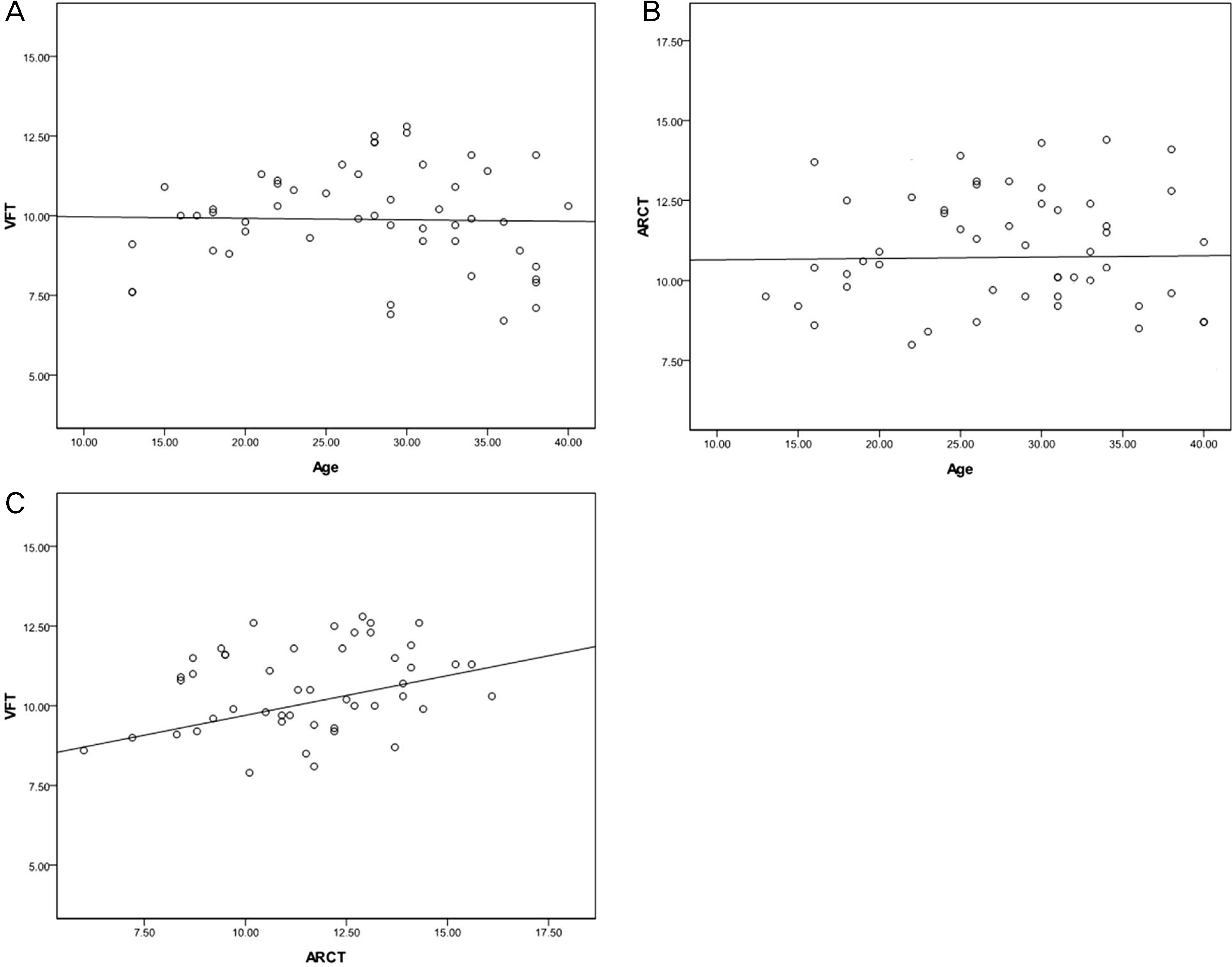
Figure 3. Graph of Spearman correlation analysis. (A) Age versus venous filling time (VFT), (B) age versus arm-retinal circulation time (ARCT), (C) arm-retinal circulation time (ARCT) versus venous filling time (VFT).
Reference
-
References
1. Novotny HR, Alvis DL. A method of photographing fluorescencein circulating blood in the human retina. Circulation. 1961; 24:82–6.2. Blair NP, Feke GT, Morales-Stoppello J, et al. Prolongation of the retinal mean circulation time in diabetes. Arch Ophthalmol. 1982; 100:764–8.
Article3. Soeldner JS, Christacopoulos PS, Gleason RE. Mean retinal circulation time as determined by fluorescein angiography in normal, prediabetic, and chemical-diabetic subjects. Diabetes. 1976; 25:903–8.4. Yang YS, Kang P, Hwang JY, Kim JD. A study on microcirculation time including retinal periphery in diabetic retinopathy using the fluorescein angiography. J Korean Ophthalmol Soc. 2000; 41:931–7.5. Sinclair SH. Macular retinal capillary hemodynamics in diabetic patients. Ophthalmology. 1991; 98:1580–6.
Article6. Wolf S, Arend O, Toonen H, et al. Retinal capillary blood flow measurement with a scanning laser ophthalmoscope. Preliminary results. Ophthalmology. 1991; 98:996–1000.
Article7. Asdourian GK, Goldberg MF. The angiographic pattern of the peripheral retinal vasculature. Arch ophthalmol. 1979; 97:2316–8.
Article8. Spitznas M, Bornfeld N. The architecture of the most peripheral retinal vessels. Albrecht von Graefes Arch kiln Exp Ophthalmol. 1977; 203:217–29.
Article9. Kang HR, Yang YS. Comparison of venous filling times and SLO findings at each quadrant region in diabetic retinopathy. Korean J Ophthalmol. 2003; 17:133–9.
Article10. Azad RV, Baishya B, Pal N, et al. Comparative evaluation of oral fluorescein angiography using the confocal scanning laser ophthalmoscope and digital fundus camera with intravenous fluorescein angiography using the digital fundus camera. Clin Experiment Ophthalmol. 2006; 34:425–9.
Article11. Tsui I, Bajwa A, Franco-Cardenas V, et al. Peripheral fluorescein angiographic findings in fellow eyes of patients with branch retinal vein occlusion. Int J Inflam. 2013; 2013:464127.
Article12. Rutnin U, Schepens CL. Fundus appearance in normal eyes. II. The standard peripheral fundus and developmental variations. Am J Ophthalmol. 1967; 64:840–52.13. Rutnin U, Schepens CL. Fundus appearance in normal eyes. 3. Peripheral degenerations. Am J Ophthalmol. 1967; 64:1040–62.14. Van Nerom PR, Rosenthal AR, Jacobson DR, et al. Iris angiography and aqueous photofluorometry in normal subjects. Arch Ophthalmol. 1981; 99:489–93.
Article
- Full Text Links
-
- Actions
-
Cited
- CITED
-
- Close
- Share
-
- Similar articles
-
- Fluorescein Angiographic Findings and Recurrence of Human Leukocyte Antigen-B27-Associated Anterior Uveitis
- Ultrawide-field Fluorescein Angiography for Evaluation of Diabetic Retinopathy
- Analysis of Therapeutic Response to Adalimumab in Behçet’s Uveitis by Ultra-Widefield Fluorescein Angiography
- Fluorescein Fundus Angiographic Findings in Diabetics
- Bilateral Fundus Findings Using Examination Under Anesthesia in Patients Showing Vitreoretinopathy at Unilateral Posterior Pole
