A Case of Canaliculocele Treated with Punctoplasty and Marsupialization
- Affiliations
-
- 1Department of Ophthalmology, Hallym University Sacred Heart Hospital, Hallym University College of Medicine, Anyang, Korea. tweeti2@hanmail.net
- 2Department of Pathology, Hallym University Sacred Heart Hospital, Hallym University College of Medicine, Anyang, Korea.
Abstract
- PURPOSE
Canaliculocele is a rare cause of eyelid mass which is formed by dilation of the canaliculus. We introduce a case of canaliculocele treated with punctoplasty and marsupialization.
CASE SUMMARY
A 35-year-old woman visited our clinic complaining of a right medial upper eyelid mass. It started 3 months ago and had the wax and wane feature. On slit lamp examination, cystic lesion was visible in the medial area of the right upper eyelid, and the punctum was obscure. Right upper canaliculus was not shown in dacryocystography. One-snip punctoplasty was performed for the diagnosis, and turbid contents were drained leading to collapse of the cyst. After diagnosis of canaliculocele, marsupialization was added by excision of the posterior wall of the mass. On histopathologic examination, the cystic wall was composed of nonkeratinized squamous epithelium with an attenuated superficial cell layer. The patient healed without recurrence of a cyst after 6 months of follow up.
Figure
-
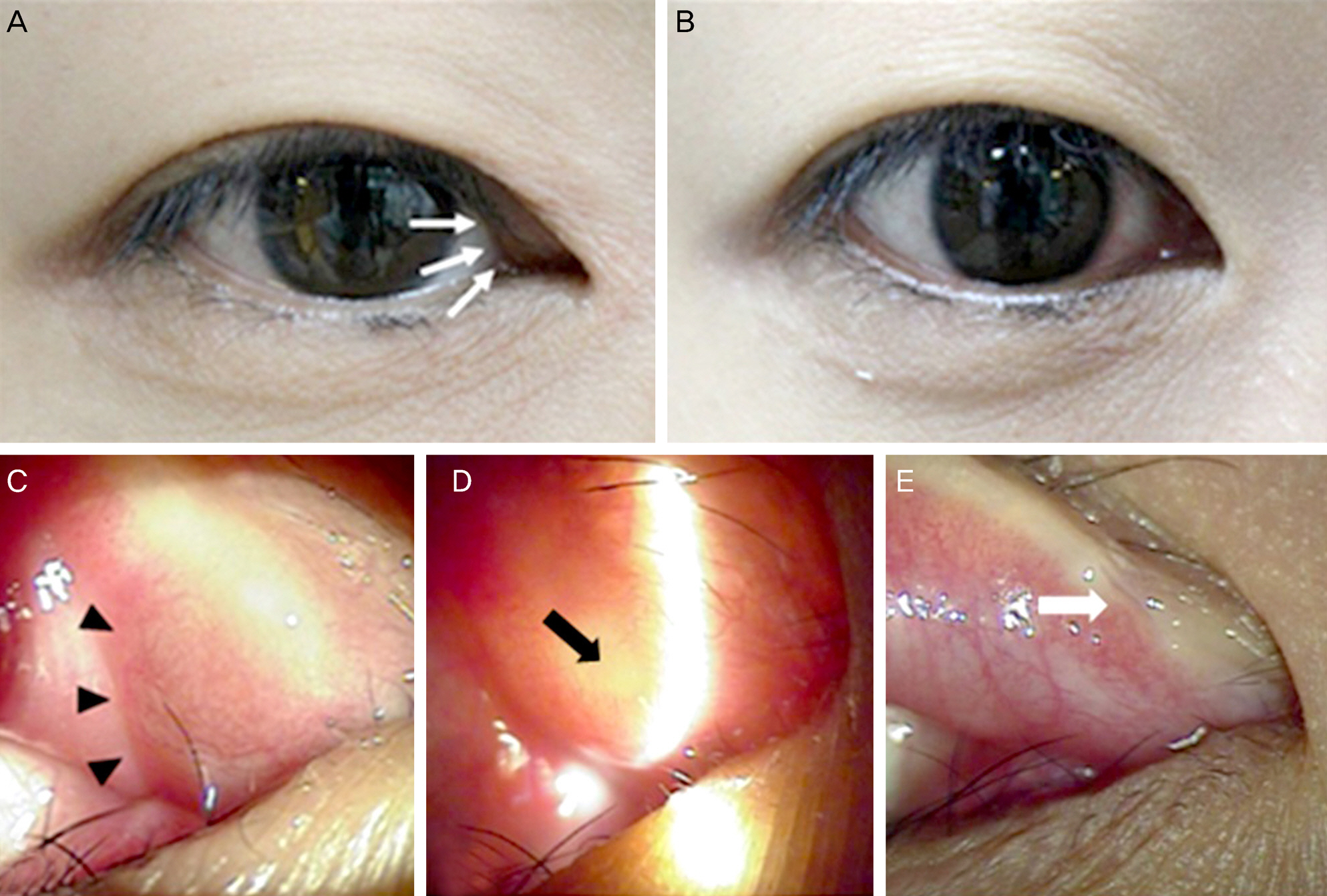
Figure 1. (A) External photograph at initial visit shows a bulging mass at the medial part of right upper eyelid (white arrows). (B) At 1month after surgery, there is no evidence of recurrence. (C) Anterior segment photograph shows a mass at infrapunctal subcon-junctival area (black arrow heads). (D) The mass is well-transilluminated with silt beam and punctal obstruction is also visible (black arrow). (E) Postoperative 6 months, the cyst is not recurred and punctual opening is well visible (white arrow).

Figure 2. Microscopic appearance of canaliculocele. (A) Longitudinal portion of canaliculocele wall. The cystic wall is composed of nonkeratinizing squamous epithelium with attenuated and flat superficial layer. The basilar germinal cells preserve the character-istic picket fence regimentation (Hematoxylin-eosin, x400). (B) Cytokeratin 7 stains the superficial layer of the epithelium (x400). (C) Cytokeratin 20 is totally negative (x400).
Reference
-
References
1. Fouad AR. A rare case of canaliculocele. Bull Ophthalmol Soc Egypt. 1972; 65:265–7.2. Sacks E, Jakobiec FA, Dodick J. Canaliculops. Ophthalmology. 1987; 94:78–81.
Article3. Kim JC, Ko YH, Woo KI, Kim YD. Canaliculocele presenting as a medial canthal mass. Ophthal Plast Reconstr Surg. 2009; 25:236–8.
Article4. Jakobiec FA, Zakka FR, Perry LP. The cytologic composition of dacryops: an immunohistochemical investigation of 15 lesions compared to the normal lacrimal gland. Am J Ophthalmol. 2013; 155:380–96. e1.
Article5. Yoon MK, Jakobiec FA, Mendoza PR. Canaliculops: clinicopatho-logic features and treatment with marsupialization. Am J Ophthalmol. 2013; 156:1062–8. e1.
Article6. Tabbara KF, Bobb AA. Lacrimal system complications in trachoma. Ophthalmology. 1980; 87:298–301.
Article7. Weatherhead RG. Wolfring dacryops. Ophthalmology. 1992; 99:1575–81.
Article8. Freedman JR, Markert MS, Cohen AJ. Primary and secondary lac-rimal canaliculitis: a review of literature. Surv Ophthalmol. 2011; 56:336–47.
Article9. Doucet TW, Hurwitz JJ. Canaliculodacryocystorhinostomy in the treatment of canalicular obstruction. Arch Ophthalmol. 1982; 100:306–9.
Article
- Full Text Links
-
- Actions
-
Cited
- CITED
-
- Close
- Share
-
- Similar articles
-
- The Effect of Punctoplasty with Pigtail Probe for Canalicular Intubation in Patients with Punctal Obstruction
- A Simple Test for Epiphora Caused by Punctal Stenosis
- The Comparison of Punctoplasty and Silicone Tube Intubation in Patients with Punctal Obstruction
- A Case of Conjunctival Inclusion Cyst Managed with Marsupialization
- Treatment of unicystic ameloblastoma by intraoral approach after marsupialization: A case report
