Clinical Evaluation of Lacrimal Gland Ductal Disease
- Affiliations
-
- 1Department of Ophthalmology, Wonkwang University School of Medicine, Institute of Wonkwang Medical Science, Iksan Korea. sangduck@wonkwang.ac.kr
Abstract
- PURPOSE
To investigate the clinical characteristics and management of lacrimal gland ductal disease, a rare disease often mistaken for other diseases.
METHODS
A retrospective chart review of 11 patients (11 eyes, 5 males, 6 females) diagnosed with lacrimal ductal disease between March 2007 and April 2013 was performed.
RESULTS
Among 11 eyes in 11 patients, 4 were diagnosed with dacryops and 7 with lacrimal gland ductulitis initiated by dacryolith. The mean age of the subjects was 47.9 years (range, 30-80 years). Lacrimal gland ductulitis patients received treatment for conjunctivitis or hordeolum for several months. Four cases involved the right eye and 7 cases involved the left eye. Symptoms included foreign body sensation, pus-like discharge and palpable mass. Biopsy was performed in 3 cases and showed no specific findings. Patients with dacryops underwent marsupialization, whereas patients diagnosed with lacrimal gland ductulitis underwent excision and dacryolith curettage. During the 2-month follow-up period, all cases showed no signs of recurrence or complications.
CONCLUSIONS
Lacrimal gland ductal disease can be mistaken for other diseases such as conjunctivitis, hordeolum, or orbital cyst, thus requiring accurate diagnosis and appropriate management.
MeSH Terms
Figure
-
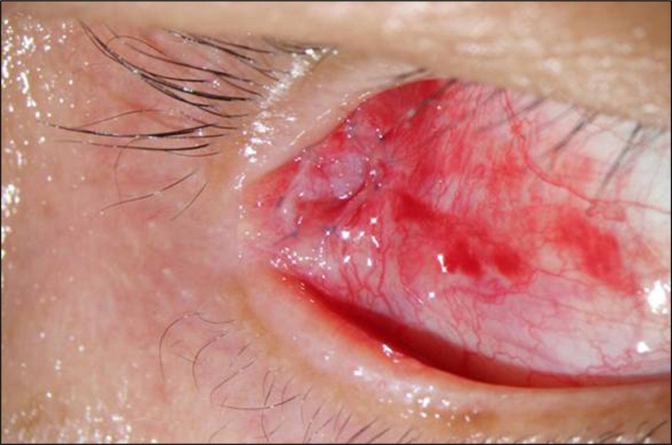
Figure 1. Post-operative photograph of marsupialization of right dacryops of 38-year-old man. The cyst walls marsu-pialized and the edges of the cyst lining were coapted to the edges of conjunctiva with 8-0 vicryl sutures.
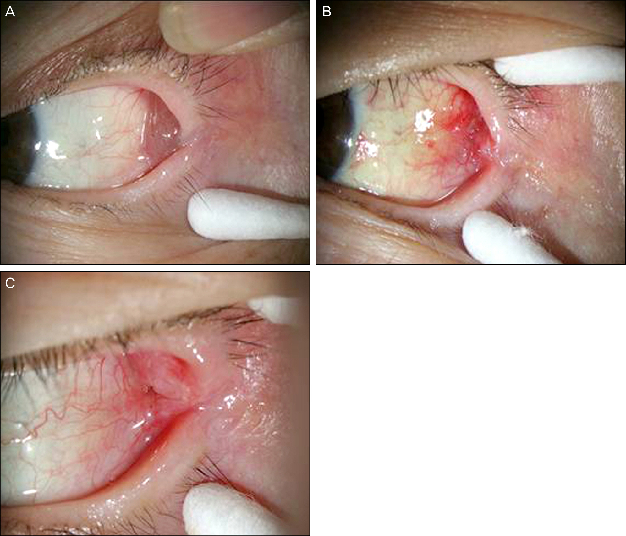
Figure 2. (A) Pre-operative photograph of dacryops of left lacrimal gland of 66-year-old female presenting with cystic mass at the left superotemporal conjunctival cul-de-sac. (B) Post-oper-ative (3 days after marsupialization) photograph. (C) Post-oper-ative (1 month after marsupialization) photograph.
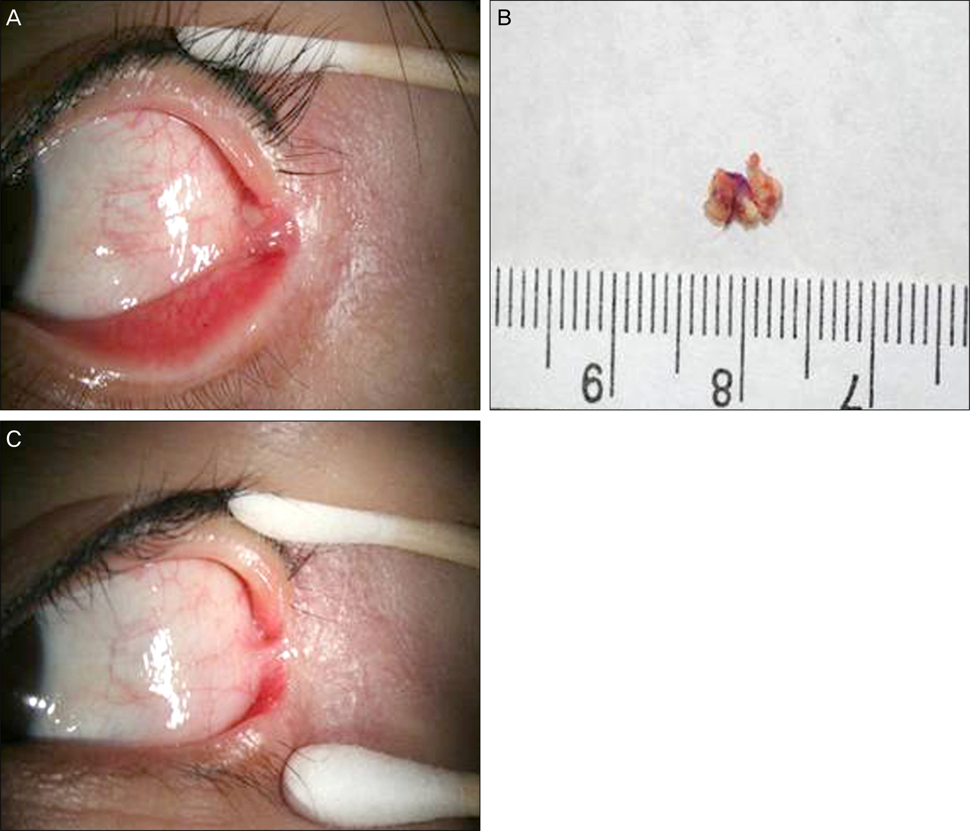
Figure 3. (A) Pre-operative photograph of a cystic mass with concretions at the left lateral canthal angle illustrated in 30-year-old female. (B) Macroscopic view of dacryolith and hair removed from the lacrimal gland ductule during the operation. (C) Post-operative (1 month after excision and dacryolith curettage) photograph.
Reference
-
References
1. Bay SW, Roh JH.Clinical characteristic of lacrimal ductal cyst. J Korean Ophthalmol Soc. 2001; 42:803–9.2. Bullock JD, Fleishman JA, Rosset JS.Lacrimal ductal cysts. Ophthalmology. 1986; 93:1355–60.
Article3. Salam A, Barrett AW, Malhotra R, Olver J.Marsupialization for lacrimal ductular cysts (dacryops): a case series. Ophthal Plast Reconstr Surg. 2012; 28:57–62.4. Harris GJ.Marsupialization of a lacrimal gland cyst. Ophthalmic Surg. 1983; 14:75–8.
Article5. Smith S, Rootman J.Lacrimal ductal cysts. Presentation and management. Surv Ophthalmol. 1986; 30:245–50.
Article6. Hornblass A, Herschorn BJ.Lacrimal gland duct cysts. Ophthalmic Surg. 1985; 16:301–6.
Article7. Park JH, Chun JS, Moon NJ, Beun DS.A case of lacrimal gland duct cyst. J Korean Ophthalmol Soc. 1994; 35:1476–80.8. Lee SH, Lew H, Yun YS.A case of lacrimal ductal cyst with dacryolith. J Korean Ophthalmol Soc. 2004; 45:131–4.9. Jeung YW, Rho JH.A case of lacrimal gland duct cyst associated with ptosis. J Korean Ophthalmol Soc. 1997; 38:1072–6.10. Zafar A, Jordan DR, Brownstein S, Faraji H.Asymptomatic lac-rimal ductule dacryolithiasis with embedded cilia. Ophthal Plast Reconstr Surg. 2004; 20:83–5.
Article11. Baker RH, Bartley GB.Lacrimal gland ductule stones. Ophthalmology. 1990; 97:531–4.
Article12. Kwon YD, Lim JW.An eyelash-induced lacrimal gland stone. J Korean Ophthalmol Soc. 2010; 51:1139–41.
Article13. Halborg J, Prause JU, Toft PB. . Stones in the lacrimal gland: a rare condition. Acta Ophthalmol. 2009; 87:672–5.
Article14. Bron AJ.Lacrimal streams: the demonstration of human lacrimal fluid secretion and the lacrimal ductules. Br J Ophthalmol. 1986; 70:241–5.
Article15. Duke-Elder S.System of ophthalmology. Part II. The ocular adnexa. Lacrimal, orbital and para-orbital diseases. 1st ed.13. London: Henry Kimpton;1974; 638–43.
Article16. Weatherhead RG.Wolfring dacryops. Ophthalmology. 1992; 99:1575–81.
Article17. Woo KI, Kim YD.Cyst of accessory lacrimal gland. Korean J Ophthalmol. 1995; 9:117–21.
Article18. Kacerovska D, Michal M, Ricarova R. . Apocrine secretion in lacrimal gland cysts (dacryops): a common but underrecognized phenomenon. J Cutan Pathol. 2011; 38:720–3.
Article19. Baratz KH, Bartley GB, Campbell RJ, Garrity JA.An eyelash nidus for dacryoliths of the lacrimal excretory and secretory systems. Am J Ophthalmol. 1991; 111:624–7.
Article20. Jakobiec FA, Zakka FR, Perry LP.The cytologic composition of dacryops: an immunohistochemical investigation of 15 lesions compared to the normal lacrimal gland. Am J Ophthalmol. 2013; 155:380–96.e1.
Article21. Eifrig CW, Chaudhry NA, Tse DT. . Lacrimal gland ductal cyst abscess. Ophthal Plast Reconstr Surg. 2001; 17:131–3.
Article
