J Korean Ophthalmol Soc.
2013 Aug;54(8):1187-1192.
Comparison of White-to-White Diameters Measured by IOLMaster, Lenstar, Orbscan, and a Manual Method
- Affiliations
-
- 1Department of Ophthalmology, Gachon University Gil Medical Center, Incheon, Korea. tigerme@naver.com
Abstract
- PURPOSE
To compare and evaluate device efficacy using white-to-white (WTW) diameter measurements by IOLMaster(R), Lenstar(R), Orbscan II(R), and a manual method with anterior segment photographs in normal eyes.
METHODS
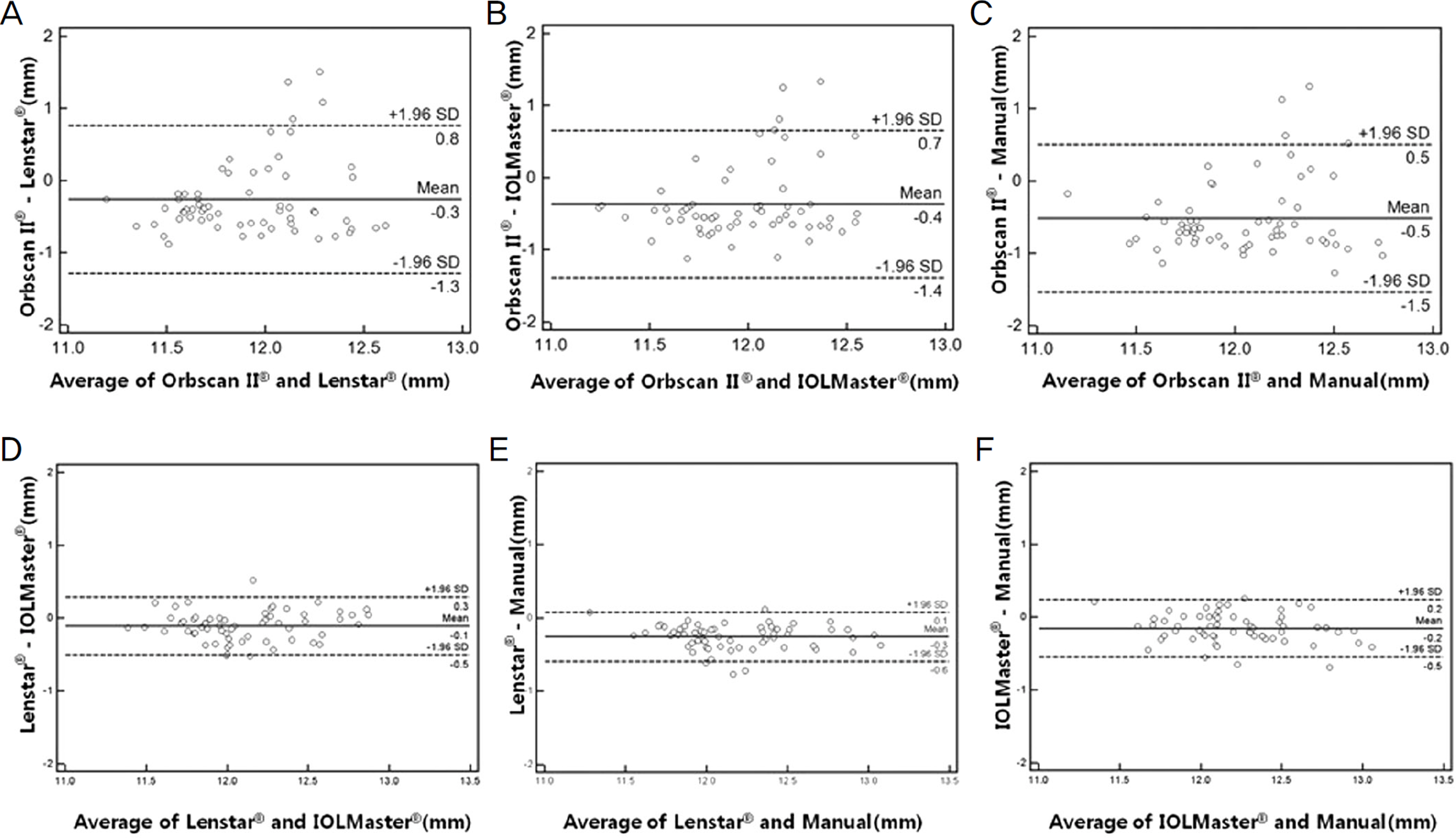
Three sets of WTW diameter measurements were obtained from 62 normal eyes of 31 patients, using the Orbscan II(R), Lenstar(R), IOLMaster(R), and a manual method with anterior segment photographs. Repeatability of each device was evaluated by coefficient of variation. ANOVA and Pearson's correlation were used to compare the differences among the devices. Bland Altman plot was performed to assess measurement agreement among the devices.
RESULTS
The mean WTW distance was 11.79 +/- 0.46 mm with Orbscan II(R), 12.05 +/- 0.38 mm with Lenstar(R), 12.15 +/- 0.36 mm with IOLMaster(R), and 12.30 +/- 0.40 mm with a manual method. There were significant differences in the results among the methods (ANOVA, p < 0.05). There were significant correlations between the devices except Orbscan II(R) (Pearson's correlation, r > 0.8, p < 0.05). The coefficient of variation of Orbscan II(R) was larger than those of Lenstar(R) and IOLMaster(R).
CONCLUSIONS
The WTW measurement using Orbscan II(R) has low correlations with other devices and lower repeatability. Our findings suggest that partial coherence interferometry should be considered as a new standard.
MeSH Terms
Figure
Reference
-
References
1. Allemann N, Chamon W, Tanaka HM. Myopic angle-supported intraocular lenses; two-year follow-up. Ophthalmology. 2000; 107:1549–54.
Article2. Gimbel HV, Ziémba SL. Management of myopic astigmatism with phakic intraocular lens implantation. J Cataract Refract Surg. 2002; 28:883–6.
Article3. Sanders DR, Schneider D, Martin R. . Toric Implantable Collamer Lens for moderate to high myopic astigmatism. Ophthalmology. 2007; 114:54–61.
Article4. Choi KH, Chung SE, Chung TY, Chung ES. Ultrasound biomicro-scopy for determining visian implantable contact lens length in-phakic IOL implantation. J Refract Surg. 2007; 23:362–7.5. Rosen E, Gore C. Staar Collamer posterior chamber phakic intra-ocular lens to correct myopia and hyperopia. J Cataract Refract Surg. 1998; 24:596–606.
Article6. Connors R 3rd, Boseman P 3rd, Olxon RJ. Accuracy and reprodu-cibility of biometry using partial coherence interferometry. J Cataract Refract Surg. 2002; 28:235–8.
Article7. Holzer MP, Mamusa M, Auffarth GU. Accuracy of a new partial coherence interferometry analyser for biometric measurements. Br J Ophthalmol. 2009; 93:807–10.
Article8. Cruysberg LP, Doors M, Verbakel F. . Evaluation of the LENSTAR LS 900 non-contact biometer. Br J Ophthalmol. 2009; 94:106–10.
Article9. Jiménez-Alfaro I, Gómez-Tellería G, Bueno JL, Puy P. Contrast sensitivity after posterior chamber phakic intraocular lens im-plantation for high myopia. J Cataract Refract Surg. 2001; 17:641–5.
Article10. Gonvers M, Bornet C, Othenin-Girard P. Implantable contact lens for moderate to high myopia: relationship of vaulting to cataract formation. J Cataract Refract Surg. 2003; 29:918–24.11. Jiménez-Alfaro I, Benítez del Castillo JM, García-Feijoó J. . Safety of posterior chamber phakic intraocular lenses for the cor-rection of high myopia: anterior segment changes after posterior chamber phakic intraocular lens implantation. Ophthalmology. 2001; 108:90–9.12. Eleftheriadis H. IOLMaster biometry: refractive results of 100 consecutive cases. Br J Ophthalmol. 2003; 87:960–3.
Article13. Vogel A, Dick HB, Krummenauer F. Reproducibility of optical biometry using partial coherence interferometry : intraobserver and interobserver reliability. J Cataract Refract Surg. 2001; 27:1961–8.14. Kielhorn I, Rajan MS, Tesha PM. . Clinical assessment of the Zeiss IOLMaster. J Cataract Refract Surg. 2003; 29:518–22.
Article15. Buckhurst PJ, Wolffsohn JS, Shah S. . A new optical low co-herence reflectometry device for ocular biometry in cataract patients. Br J Ophthalmol. 2009; 93:949–53.
Article16. Baumeister M, Terzi E, Ekici Y, Kohnen T. Comparison of manual and automated methods to determine horizontal corneal diameter. J Cataract Refract Surg. 2004; 30:374–80.
Article17. Nemeth G, Hassan Z, Szalai E. . Comparative analysis of white-to-white and angle-to-angle distance measurements with partial coherence interferometry and optical coherence tomography. J Cataract Refract Surg. 2010; 36:1862–6.
Article18. Freeman G, Pesudovs K. The impact of cataract severity on meas-urement acquisition with the IOLMaster. Acta Ophthalmol Scand. 2005; 83:439–42.
Article19. Dinc UA, Oncel B, Gorgun E. . Assessment and comparison of anterior chamber dimensions using various imaging techniques. Ophthalmic Surg Lasers Imaging. 2010; 41:115–22.
Article20. Oh J, Shin HH, Kim JH. . Direct measurement of the ciliary sulcus diameter by 35-megahertz ultrasound biomicroscopy. Ophthalmology. 2007; 114:1685–8.
Article21. Lee SC, Jin KH. Ciliary sulcus size according to refractive error using ultrasound biomicroscopy. J Korean Ophthalmol Soc. 2004; 45:2093–8.
- Full Text Links
- Actions
-
Cited
- CITED
-
- Close
- Share
- Similar articles
-
- Comparison of Anterior Segment Measurements Using Scanning-Slit Topography and Optical Low-Coherence Reflectometry (OLCR) Biometry
- Comparison of Anterior Segment Measurements with a New Multifunctional Unit and Five Other Devices
- Comparison of Ocular Biometry Using New Swept-source Optical Coherence Tomography-based Optical Biometer with Other Devices
- Efficacy of Biometry Using Swept-source Optical Coherence Tomography for Posterior Chamber Phakic Intraocular Lens Implantation
- The Accuracy of Axial Length Measurement Using Partial Coherence Interferometrys


