J Korean Ophthalmol Soc.
2012 Feb;53(2):333-337.
A Case of Acquired Dacryocystocele Treated by Lacrimal Silicone Intubation
- Affiliations
-
- 1Kong Eye Clinic, Seoul, Korea.
- 2Department of Ophthalmology, Seoul National University Hospital, Seoul, Korea. khwarg@snu.ac.kr
- 3Department of Ophthalmology, Seoul National University College of Medicine, Seoul, Korea.
Abstract
- PURPOSE
To report a case of an acquired dacryocystocele successfully treated with bicanalicular silicone intubation and to review relating literature.
CASE SUMMARY
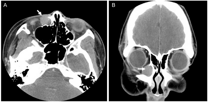
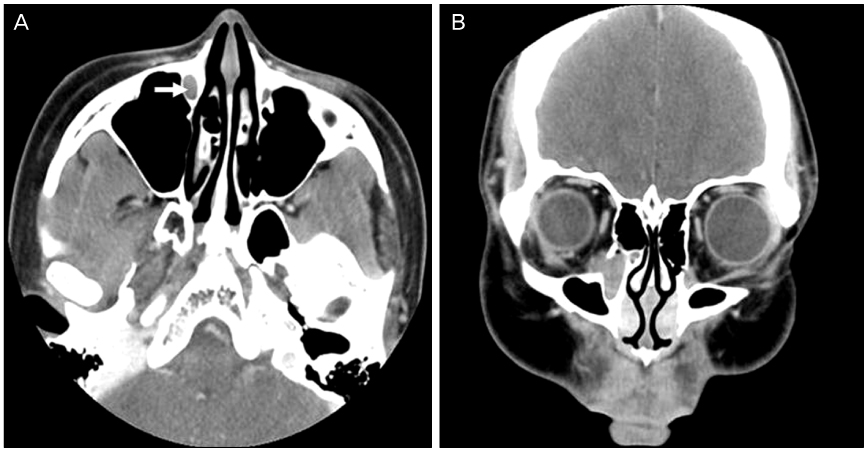
A 17-year-old girl visited our clinic with tearing of both eyes since birth and a mass on the right medial canthal area for 2 years. A firm, non-tender mass with a well-demarcated border was palpated in the subcutaneous level just inferior to the right medial canthal ligament. Lacrimal irrigation via the lower punctums showed reflux through the opposite punctums without nasal passage in both of her eyes. Computed tomographic scan showed a widening of the right lacrimal sac fossa and bony nasolacrimal canal and a 16 x 18 mm sized cyst-like mass in the right lacrimal sac. The patient was diagnosed with right acquired dacryocystocele associated with bilateral congenital nasolacrimal duct obstructions. After opening of the obstructed common canaliculus using a fine lacrimal probe, silicone intubation was performed. The tearing symptom improved and the mass disappeared during the subsequent follow-up period of 1 year.
CONCLUSIONS
When only accompanied by distal nasolacrimal duct obstruction, acquired dacryocystocele can be inferred to be associated with congenital nasolacrimal duct obstruction. Subsequently, bicanalicular silicone intubation can be considered as a treatment of choice.
MeSH Terms
Figure
Reference
-
1. Harris GJ, DiClementi D. Congenital dacryocystocele. Arch Ophthalmol. 1982. 100:1763–1765.2. On YH, Shin H. A case report of congenital dacryocystocele (Dacryocystitis Neonatorum). J Korean Ophthalmol Soc. 1984. 25:297–301.3. Mansour AM, Cheng KP, Mumma JV, et al. Congenital dacryocele. A collaborative review. Ophthalmology. 1991. 98:1744–1751.4. Lee JH, Moon SW, Shin YW, Lee YJ. Dacryocystocele in adult: A report of five cases. J Korean Ophthalmol Soc. 2010. 51:751–757.5. Jones LT, Wobig JL. Surgery of the Eyelids and Lacrimal System. 1976. 1st ed. Birmingham, AL: Aesculapius Publishing Co;162–167.6. Weinstein GS, Biglan AW, Patterson JH. Congenital lacrimal sac mucoceles. Am J Ophthalmol. 1982. 94:106–110.7. Woo KI, Kim YD. Four cases of dacryocystocele. Korean J Ophthalmol. 1997. 11:65–69.8. Lai PC, Wang JK, Liao SL. A case of dacryocystocele in an adult. Jpn J Ophthalmol. 2004. 48:419–421.9. Wong RK, VanderVeen DK. Presentation and management of congenital dacryocystocele. Pediatrics. 2008. 122:e1108–e1112.10. Schnall BM, Christian CJ. Conservative treatment of congenital dacryocele. J Pediatr Ophthalmol Strabismus. 1996. 33:219–222.11. Becker BB. The treatment of congenital dacryocystocele. Am J Ophthalmol. 2006. 142:835–838.12. Eloy P, Martinez A, Leruth E, et al. Endonasal endoscopic dacryocystorhinostomy for a primary dacryocystocele in an adult. B-ENT. 2009. 5:179–182.13. Bhaya MH, Meehan R, Har-El G. Dacryocystocele in an adult: endoscopic management. Am J Otolaryngol. 1997. 18:131–134.14. Menestrina LE, Osborn RE. Congenital dacryocystocele with intranasal extension: correlation of computed tomography and magnetic resonance imaging. J Am Osteopath Assoc. 1990. 90:264–268.15. Teixeira CC, Dias RJ, Falcão-Reis FM, Santos M. Congenital dacryocystocele with intranasal extension. Eur J Ophthalmol. 2005. 15:126–128.16. Oh YK, Lee DS, Kim SY, Ye MK. A case of bilateral congenital dacryocystoceles with nasolacrimal duct cysts. J Korean Ophthalmol Soc. 2002. 43:781–785.17. Shekunov J, Griepentrog GJ, Diehl NN, Mohney BG. Prevalence and clinical characteristics of congenital dacryocystocele. J AAPOS. 2010. 14:417–420.18. Cavazza S, Laffi GL, Lodi L, et al. Congenital dacryocystocele: diagnosis and treatment. Acta Otorhinolaryngol Ital. 2008. 28:298–301.19. Shashy RG, Durairaj V, Holmes JM, et al. Congenital dacryocystocele associated with intranasal cysts: diagnosis and management. Laryngoscope. 2003. 113:37–40.
- Full Text Links
- Actions
-
Cited
- CITED
-
- Close
- Share
- Similar articles
-
- A Case of Acquired Lacrimal Fistula Caused by Silicone Tube Remnant
- Surgical Efficacy of Probing with Silicone Intubation for Lacrimal Apparatus Obstruction in Children
- A Case of Endoscopic Management of Dacryocystocele in an Adult
- Silicone Tube Intubation with Lacrimal Endoscopy and Endonasal Dacryocystorhinostomy in Adult Nasolacrimal Duct Obstruction
- Results with Silicone Stent in Lacrimal Drainage System


