The Effectiveness of Mitomycin C on Pterygium Surgery with Amniotic Membrane Transplantation
- Affiliations
-
- 1Department of Ophthalmology, Daegu Fatima Hospital, Daegu, Korea. djoph2540@yahoo.co.kr
Abstract
- PURPOSE
To compare postoperative recurrence rates of pterygium surgery with amniotic membrane transplantation with and without mitomycin C.
METHODS
A retrospective analysis of 45 eyes of 43 patients who underwent pterygium surgery with amniotic membrane transplantation with a minimum of six months of follow-up. Nineteen eyes underwent mitomycin C application, while the remaining 26 eyes did not. Recurrence rates and complications were evaluated.
RESULTS
With a minimum of six months of follow-up, fibrovascular tissue in the excised area not invading the cornea (conjunctival recurrence) was noted in two eyes (10.5%), and fibrovascular tissue invading the cornea (corneal recurrence) was noted in one eye (5.4%) in the amniotic membrane transplantation with mitomycin C group. Conjunctival recurrence was noted in six eyes (23.1%) and corneal recurrence in three eyes (11.5%) in the amniotic membrane transplantation without mitomycin C group. Recurrence rate in the amniotic membrane transplantation with mitomycin C group (15.9%) was significantly lower (p = 0.014) than that in the amniotic membrane transplantation without mitomycin C group (34.6%). Complications included sub-amniotic membrane hemorrhage in two eyes, granuloma in one eye, and wound dehiscence in one eye in each group. There were no specific complications related to usage of mitomycin C.
CONCLUSIONS
In pterygium surgery with amniotic membrane transplantation, application of mitomycin C is an effective method to reduce recurrence rates, especially conjunctival recurrences that are related to cosmetic problems. This method may also be helpful to reduce patient discomfort.
MeSH Terms
Figure
-
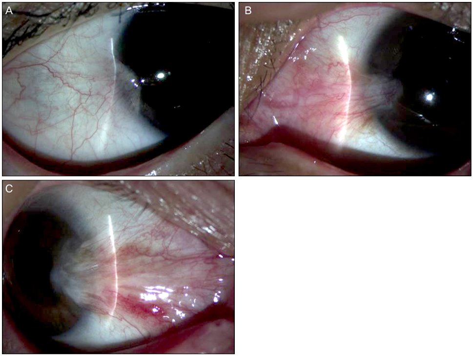
Figure 1 Classification of pterygium. (A) Grade T1 (atrophic), episcleral vessels are unobscured. (B) Grade T2 (intermediate), episcleral vessels are partially obscured. (C) Grade T3 (fleshy), episcleral vessels are totally obscured.

Figure 2 This procedure was performed as fellows; (A) Episcleral traction sutures were placed. (B) Excision and blunt dissection of pterygium from the cornea and sclera by using conjunctival scissors. (C) Remnant pterygium of the cornea was dissected off by corneal forceps. (D) Wide excision of the subconjunctival Tenon's tissue was done by the Ellman cautery. (E) 0.02% Mitomycin C soaked cotton swabs were applied at excision site. (F) Amniotic membrane is peeled off from the carrier paper and spread over the cornea. (G) The amniotic membrane was sutured to the episclera and conjuctiva to facilitate regeneration of the conjunctiva over the amniotic membrane.

Figure 3 These figures showed the two kinds of recurrent pterygium. (A, B) Grade 2, fibrovascular tissue in the excised area, reaching to the limbus, but not invading the cornea (conjunctival recurrence). (C, D) Grade 3, fibrovascular tissue invading the cornea (corneal recurrence).
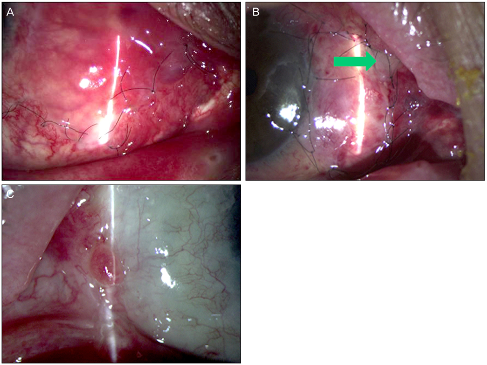
Figure 4 Complications of pterygium excision with amniotic membrane transplantation. (A) Sub-amniotic membrane hemorrhage. (B) Wound gapping (arrow). (C) Granuloma.
Reference
-
1. Dushku N, John MK, Schultz GS, Reid TW. Pterygia pathogenesis: corneal invasion by matrix metalloproteinase expressing altered limbal epithelial basal cells. Arch Ophthalmol. 2001. 119:695–706.2. Pinkerton OD, Hokama Y, Shigemura LA. Immunologic basis for the pathogenesis of pterygium. Am J Ophthalmol. 1984. 98:225–228.3. Lee SH, Jeong HJ. Immune reactions in pterygium. J Korean Ophthalmol Soc. 1987. 28:927–933.4. Bahrassa F, Datta R. Postoperative beta irradiation treatment of pterygium. Int J Radiat Oncol Biol Phys. 1983. 9:679–684.5. Kim CH, Lee JK, Park DJ. Recurrence rates of amniotic membrane transplantation, conjunctival autograft and conjunctivolimbal autograft in primary pterygium. J Korean Ophthalmol Soc. 2009. 50:1780–1788.6. Hayasaka S, Noda S, Yamamoto Y, Setogawa T. Postoperative instillation of low dose mitomycin C in the treatment of primary pterygium. Am J Ophthalmol. 1988. 106:715–718.7. Frucht-Pery J, Siganos CS, Ilsar M. Intraoperative application of topical mitomycin C for pterygium surgery. Ophthalmology. 1996. 103:674–677.8. Rubinfeld RS, Pfister RR, Stein RM, et al. Serious complications of topical mitomycin C after pterygium surgery. Ophthalmology. 1992. 99:1647–1654.9. Hirst LW. The treatment of pterygium. Surv Ophthalmol. 2003. 48:145–180.10. Prabhasawat P, Barton K, Burkett G, Tseng SC. Comparison of conjunctival autografts, amniotic membrane grafts, and primary closure for pterygium excision. Ophthalmology. 1997. 104:974–985.11. Solomon A, Pires RT, Tseng SC. Amniotic membrane transplantation after extensive removal of primary and recurrent pterygia. Ophthalmology. 2001. 108:449–460.12. Jang JH, Choi TH. The effect of amniotic membrane transplantation for pterygium excision. J Korean Ophthalmol Soc. 2005. 46:597–604.13. Lee BH, Lee JW, Park YJ, Lee KW. Clinical research on effectiveness of mitomycin C on primary pterygium with limbal-conjunctival autograft. J Korean Ophthalmol Soc. 2009. 50:996–1004.14. Tan DT, Chee SP, Dear KB, Lim AS. Effect of pterygium morphology on pterygium recurrence in a controlled trial comparing conjunctival autografting with bare sclera excision. Arch Ophthalmol. 1997. 115:1235–1240.15. Mutlu FM, Sobaci G, Tatar T, Yildirim E. A comparative study of recurrent pterygium surgery: limbal conjunctival autograft transplantation versus mitomycin C with conjunctival flap. Ophthalmology. 1999. 106:817–821.16. Verweij J, Pinedo HM. Mitomycin C: mechanism of action, usefulness and limitations. Anticancer Drugs. 1990. 1:5–13.17. Kunitomo N, Mori S. Studies on the pterygium. Report IV. A treatment of the pterygium by mitomycin C instillation. Nippon Ganka Gakkai Zasshi. 1963. 67:601–607.18. van Herendael BJ, Oberti C, Brosens I. Microanatomy of the human amniotic membrane. A light microscopic, transmission, and scanning electron microscopic study. Am J Obstet Gynecol. 1978. 131:872–880.19. Terranova VP, Lyall RM. Chemotaxis of human gingival epithelial cells to laminin. A mechanism for epithelial cell apical migration. J Periodontol. 1986. 57:311–317.20. Sonnenberg A, Calafat J, Janssen H, et al. Integrin alpha 6/beta 4 complex is located in hemidesmosomes, suggesting a major role in epidermal cell-basement membrane adhesion. J Cell Biol. 1991. 113:907–917.21. Streuli CH, Bailey N, Bissel MJ. Control of mammary epithelial differentiation: basement membrane induces tissue-specific gene expression in the absence of cell-cell interaction and morphological polarity. J Cell Biol. 1991. 115:1383–1395.22. Na BK, Hwang JH, Kim JC, et al. Analysis of human amniotic membrane components as proteinase inhibitors for development of therapeutic agent for recalcitrant keratitis. Placenta. 1999. 20:453–466.23. Ti SE, Tseng SC. Management of primary and recurrent pterygium using amniotic membrane transplantation. Curr Opin Ophthalmol. 2002. 13:204–212.24. Tananuvat N, Martin T. The results of amniotic membrane transplantation for primary pterygium compared with conjunctival autograft. Cornea. 2004. 23:458–463.
- Full Text Links
-
- Actions
-
Cited
- CITED
-
- Close
- Share
-
- Similar articles
-
- Ocular Surface Reconstruction with Amniotic Membrane Transplantation in Pterygium
- The Effect of Amniotic Membrane Transplantation for Pterygium Excision
- Recurrence Rates of Conjunctival Autograft Transplantation With Aminiotic Membrane Transplantation in Primary Pterygium Surgery
- Surgical Outcome of Primary Pterygium Excision with Conjunctival Autograft
- Amniotic Membrane Transplantation and Use of Adjunctive Mitomycin C in the Treatment of Symblepharon
