J Korean Ophthalmol Soc.
2011 Apr;52(4):507-510.
Transient Visual Loss in Peripapillary Staphyloma
- Affiliations
-
- 1Department of Ophthalmology, Chungnam National University College of Medicine, Daejeon, Korea. opticalyh@hanmail.net
- 2Chungnam National University Research Institute for Medical Sciences, Daejeon, Korea.
Abstract
- PURPOSE
To report the case of a patient with transient visual loss with a peripapillary staphyloma.
CASE SUMMARY
The authors of the present study examined a 30-year-old woman who complained of transient visual loss in her right eye. The patient lost her vision for 5 seconds on average approximately 5 times a day. Her visual acuity was 20/20 in both eyes. There were no abnormalities on slit lamp examination. A deep excavation with choroidal atrophy in the peripapillary area of the right eye was found. The patient was diagnosed with peripapillary staphyloma. Her physiological blind spot in the right eye was enlarged on Humphrey visual field testing. However, she had no abnormalities on color vision testing, fluorescein angiography, or magnetic resonance imaging (MRI). There were no abnormalities in the evaluation of the brain, carotid arteries, or heart. There was no evidence of vasculitis or hypercoagulability. The symptom did not change on pressing the eyeball, performing the Valsalva maneuver or carotid massage, applying cycloplegics, or shining a strong light in the other eye. The symptom did not change after taking a calcium channel blocker for 4 weeks.
Keyword
MeSH Terms
Figure
-
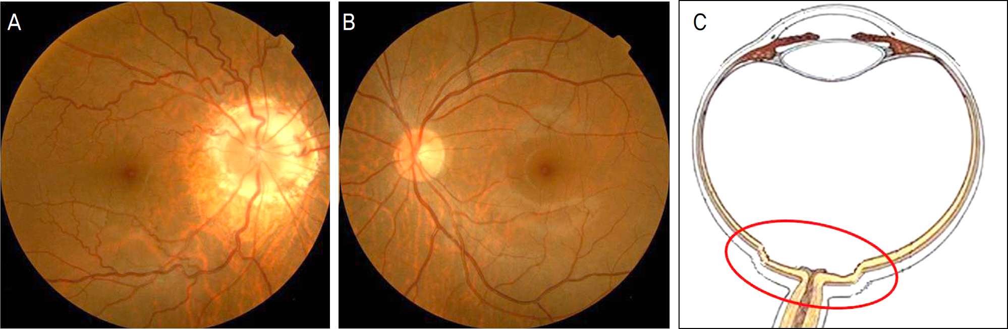
Figure 1. (A) Fundus photograph of the right eye shows a deep excavation on the peripapillary area with attenuated choroidal pigment and pigmented epithelium. Normal-appearing optic disc is centrally located in the excavation. (B) Fundus photograph of the normal left eye. (C) Schematic drawing of peripapillary staphyloma.

Figure 2. Enlarged physiologic blind spot in the right eye on Humphrey visual field test.
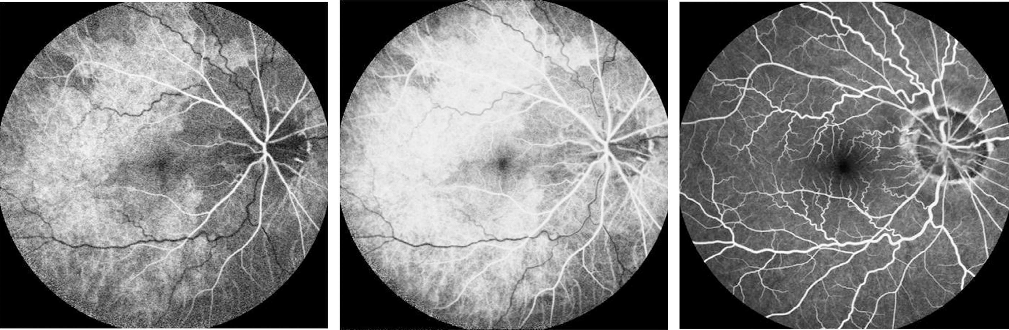
Figure 3. Fluorescein angiographs show normal findings except peripapillary abnormality by peripapillary staphyloma.
Reference
-
References
1. Current management of amaurosis fugax. The Amaurosis Fugax Study Group. Stroke. 1990; 21:201–8.2. Miller FW, Santoro TJ. Nifedipine in the treatment of migraine headache and amaurosis fugax in patients with systemic lupus erythematosus. N Engl J Med. 1984; 311:921.
Article3. Shaw HE Jr, Osher RH, Smith JL. Amaurosis fugax associated with SC hemoglobinopathy and lupus erythematosus. Am J Ophthalmol. 1979; 87:281–5.
Article4. Brown G, Tasman W. Congenital Anomalies of the Optic Disc. New York: Grune&Stratton;1983. p. 95–191.5. Brodsky MC. Congenital optic disk anomalies. Surv Ophthalmol. 1994; 39:89–112.
Article6. Brodsky MC, Baker RS, Hemed LM. Pediatric Neuro-Ophthalmology. New York: Springer-Verlag;1996. p. 49–56.7. Miller NR. Walsh and Hoyt's Clinical Neuro-Ophthalmology. 6th ed.1. Baltimore: Lippincott Williams & Wilkins;2005. p. 159–65.8. Meirelles RL, Aggio FB, Costa RA, Farah ME. STRATUS optical coherence tomography in unilateral colobomatous excavation of the optic disc and secondary retinoschisis. Graefes Arch Clin Exp Ophthalmol. 2005; 243:76–81.
Article9. Srinivasan G, Venkatesh P, Garg S. Optical coherence tomographic characteristics in morning glory disc anomaly. Can J Ophthalmol. 2007; 42:307–9.
Article10. Sugar HS, Beckman H. Peripapillary staphyloma with respiratory pulsation. Am J Ophthalmol. 1969; 68:895–7.11. Kral K, Svarc D. Contractile peripapillary staphyloma. Am J Ophthalmol. 1971; 71:1090–2.
Article12. Burger SK, Saul RF, Selhorst JB, Thurston SE. Transient monocular blindness caused by vasospasm. N Engl J Med. 1991; 325:870–3.
Article13. Jehn A, Frank Dettwiler B, Fleischhauer J, et al. Exercise-induced vasospastic amaurosis fugax. Arch Ophthalmol. 2002; 120:220–2.14. Miller NR. Walsh and Hoyt's Clinical Neuro-Ophthalmology. 6th ed.2. Baltimore: Lippincott Williams & Wilkins;2005. p. 2035–6.15. Graether JM. Transient amaurosis in one eye with simultaneous di-latation of retinal veins. In Association with a congenital anomaly of the optic nerve head. Arch Ophthalmol. 1963; 70:342–5.16. Liu GT. Neuro-Ophthalmology: Diagnosis and management. Saunders;2000. p. 379–80.17. Seybold ME, Rosen PN. Peripapillary staphyloma and amaurosis fugax. Ann Ophthalmol. 1977; 9:1139–41.
- Full Text Links
-
- Actions
-
Cited
- CITED
-
- Close
- Share
-
- Similar articles
-
- A Case of Retinal Detachment with Equatorial Scleral Staphyloma
- A Case of Retinal Detachment with Equatorial Scleral Staphyloma
- Transient Visual Loss after Retrobulbar Anesthesia in the Advanced Glaucoma Patients
- Comparison of Optic Ne rve Head Blood Flow in Normal-tension Glaucoma with Asymmetric Visual Field Loss
- Two Cases of Intrapapillary Hemorrhage with Adjacent Peripapillary Subretinal Hemorrhage
