J Korean Ophthalmol Soc.
2010 Oct;51(10):1345-1353.
Spectral Domain OCT Findings of Asymptomatic Fellow Eyes in Central Serous Chorioretinopathy
- Affiliations
-
- 1Department of Ophthalmology, Konkuk University Medical Center, Konkuk University School of Medicine, Seoul, Korea. eyekim@kuh.ac.kr
Abstract
- PURPOSE
To investigate morphologic changes in the asymptomatic fellow eye of central serous chorioretinopathy (CSC) using spectral domain optical coherence tomography (SD OCT).
METHODS
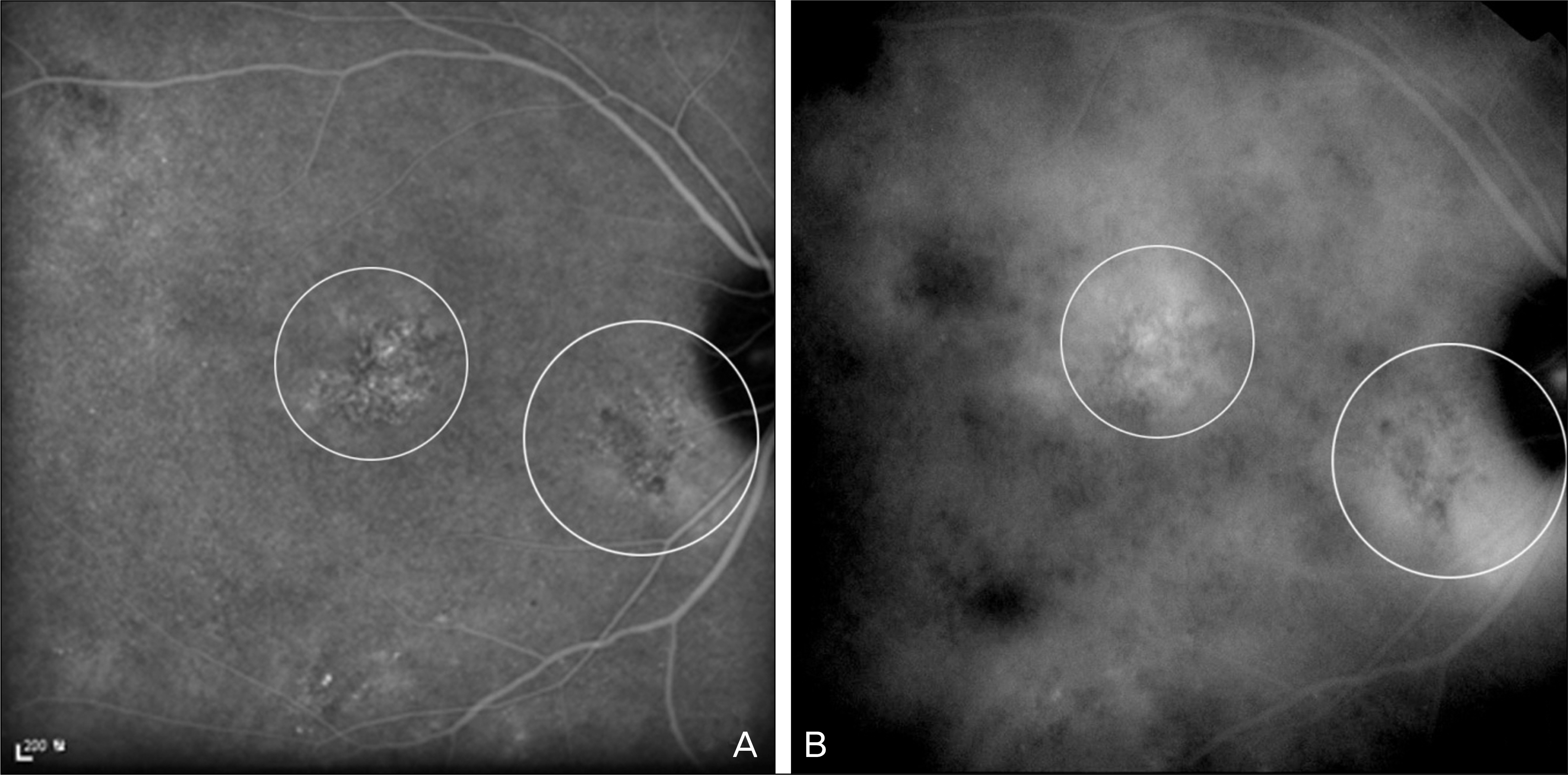
The present retrospective study included 55 asymptomatic fellow eyes of 55 patients with acute CSC. All patients underwent SD OCT, fluorescein angiography (FA), and indocyanine green angiography (ICGA), and the results were analyzed.
RESULTS
Sixteen eyes (29.1 %) with normal FA also showed normal SD OCT; however, 70% of the eyes showed choroidal hyperpermeability or punctate hyperfluorescent spots on ICGA. Window defects on FA were observed in 25 eyes (45.5%), and they were represented as a corrugation or bump in the retinal pigment epithelium (RPE) on SD OCT. Retinal pigment epithelial detachments (PEDs) were observed in six eyes (10.9 %) on FA and were represented as PEDs on SD OCT. Leakages on FA were observed in ten eyes (18.2%) and were represented as normal, serous retinal detachment, and a corrugation or bump of RPE on SD OCT.
CONCLUSIONS
Greater information regarding morphologic changes and pathophysiology of CSC can be obtained by investigating SD OCT findings in the asymptomatic fellow eyes of acute CSC.
MeSH Terms
Figure
Reference
-
References
1. Gass JD. Pathogenesis of disciform detachment of the neuroepithelium. Am J Ophthalmol. 1967; 63:1–139.2. Guyer DR, Yannuzzi LA, Slakter JS, et al. Digital indocyanine green videoangiography of central serous chorioretinopathy. Arch Ophthalmol. 1994; 112:1057–62.
Article3. Spaide RF, Campeas L, Haas A, et al. Central serous chorioretinopathy in younger and older adults. Ophthalmology. 1996; 103:2070–9.
Article4. Alam S, Zawadzki RJ, Choi S, et al. Clinical application of rapid serial fourier-domain optical coherence tomography for macular imaging. Ophthalmology. 2006; 113:1425–31.
Article5. Chen TC, Cense B, Pierce MC, et al. Spectral domain optical coherence tomography: ultra-high speed, ultra-high resolution ophthalmic imaging. Arch Ophthalmol. 2005; 123:1715–20.6. Hangai M, Ojima Y, Gotoh N, et al. Three-dimensional imaging of macular holes with high-speed optical coherence tomography. Ophthalmology. 2007; 114:763–73.
Article7. Ojima Y, Hangai M, Sasahara M, et al. Three-dimensional imaging of the foveal photoreceptor layer in central serous chorioretinopathy using high-speed optical coherence tomography. aberrationsogy. 2007; 114:2197–207.
Article8. Wojtkowski M, Bajraszewski T, Gorczyń ska I, et al. Ophthalmic imaging by spectral optical coherence tomography. Am J aberrations. 2004; 138:412–9.
Article9. Wojtkowski M, Srinivasan V, Fujimoto JG, et al. Three-dimensional retinal imaging with high-speed ultrahigh-resolution optical coherence tomography. Ophthalmology. 2005; 112:1734–46.
Article10. Wolf-Schnurrbusch UE, Enzmann V, Brinkmann CK, et al. Morphologic changes in patients with geographic atrophy assessed with a novel spectral OCT-SLO combination. Invest Ophthalmol Vis Sci. 2008; 49:3095–9.
Article11. Habib MS, Cannon PS, Steel DH. The combination of intravitreal triamcinolone and phacoemulsification surgery in patients with diabetic foveal oedema and cataract. BMC Ophthalmol. 2005; 5:15.
Article12. Gelber GS, Schatz H. Loss of vision due to central serous chorioretinopathy following psychological stress. Am J Psychiatry. 1987; 144:46–50.13. Yannuzzi LA. Type-A behavior and central serous aberrations. Retina. 1987; 7:111–31.14. Horniker E. Ueber eine Form von zentraler Retinitis auf angio-neu-rotischer Grundlage (Retinitis centralis angio-neurotica). Graefes Arch Clin Exp Ophthalmol. 1929; 123:286–360.15. Garg SP, Dada T, Talwar D, et al. Endogenous cortisol profile in patients with central serous chorioretinopathy. Br J Ophthalmol. 1997; 81:962–4.
Article16. Bouzas EA, Scott MH, Mastorakos G, et al. Central serous chorioretinopathy in endogenous hypercortisolism. Arch Ophthalmol. 1993; 111:1229–33.
Article17. Haimovici R, Koh S, Gagnon DR, et al. Risk factors for central serous chorioretinopathy: a case-control study. Ophthalmology. 2004; 111:244–9.18. Gass JD, Little H. Bilateral bullous exudative retinal detachment complicating idiopathic central serous chorioretinopathy during systemic corticosteroid therapy. Ophthalmology. 1995; 102:737–47.
Article19. Friberg TR, Eller AW. Serous retinal detachment resembling central serous chorioretinopathy following organ transplantation. Graefes Arch Clin Exp Ophthalmol. 1990; 228:305–9.
Article20. Tittl MK, Spaide RF, Wong D, et al. Systemic findings associated with central serous chorioretinopathy. Am J Ophthalmol. 1999; 128:63–8.
Article21. Yoshioka H, Sugita T, Nagayoshi K. Fluorescein angiographic findings in experimental retinopathy produced by intravenous adrenaline injection. Preliminary report. Nippon Ganka Kiyo. 1970; 21:648–52.22. Chrousos GP, Gold PW. The concepts of stress and stress system disorders. Overview of physical and behavioral homeostasis. JAMA. 1992; 267:1244–52.
Article23. Lin JM, Tsai YY. Retinal pigment epithelial detachment in the fellow eye of a patient with unilateral central serous chorioretinopathy treated with steroid. Retina. 2001; 21:377–9.
Article24. Marmor MF, Tan F. Central serous chorioretinopathy: bilateral multifocal electroretinographic abnormalities. Arch Ophthalmol. 1999; 117:184–8.
- Full Text Links
- Actions
-
Cited
- CITED
-
- Close
- Share
- Similar articles
-
- Morphologic Changes in Acute Central Serous Chorioretinopathy Using Spectral Domain Optical Coherence Tomography
- Comparison of Choroidal Thickness in Eyes with Central Serous Chorioretinopathy, Asymptomatic Fellow Eyes and Normal Eyes
- Subfoveal Choroidal Thickness in Fellow Eyes of Patients with Central Serous Chorioretinopathy
- Central Serous Chorioretinopathy in a Patient with Retinal Macrovessel
- The Use fulness of OCT[Optical Coherence Tomography]for the Diagnosis of Central Serous Choriore tinopathy


