J Korean Ophthalmol Soc.
2010 Aug;51(8):1155-1160.
Anterior Ischemic Optic Neuropathy Following Periocular Autologous Fat Injection
- Affiliations
-
- 1Department of Ophthalmology, Gachon University Gil Hospital, Incheon, Korea. ldy90@hanmail.net
Abstract
- PURPOSE
To report an unusual case of anterior ischemic optic neuropathy and choroidal ischemia after injection of autologous fat into the periorbital region due to embolism of the short posterior ciliary artery without involving the retinal artery.
CASE SUMMARY
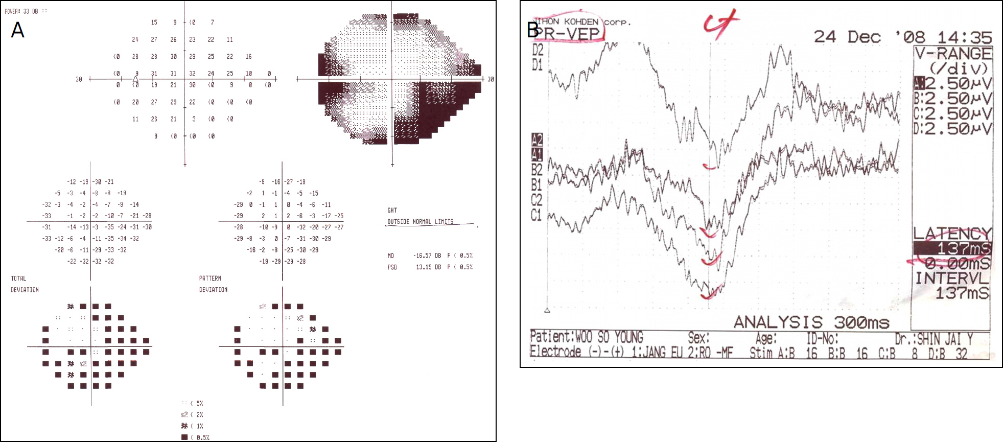
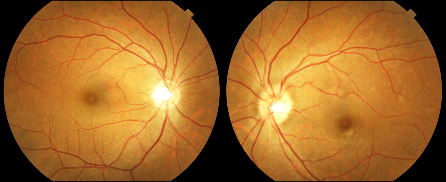
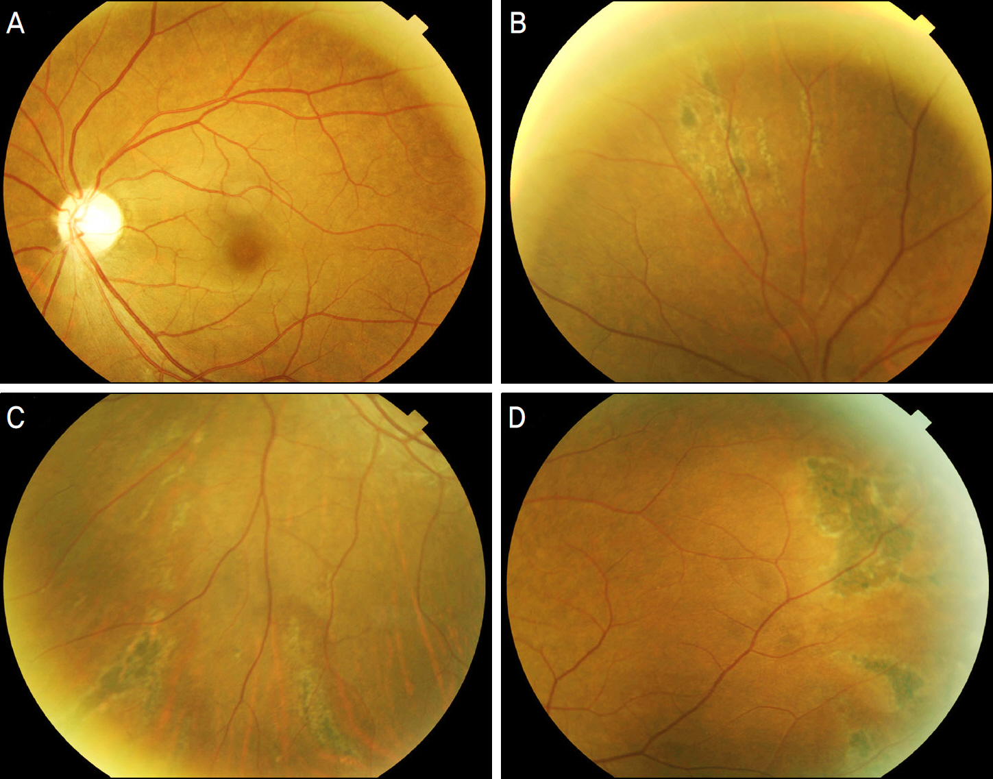
A 39-year-old female presented with sudden blurred vision, diplopia and ptosis of her left eye immediately after receiving an autologous fat injection into the periorbital area. The first ophthalmologic examination revealed that the patient s left eye had decreased visual acuity, relative afferent pupillary defect, exotropia, and hypertropia. Fundus examination of the left eye showed disc edema. Fluorescein angiography showed multiple choroidal vascular filling defects at the early phase and wedge-shaped or geographic fluorescein staining at the superior, inferior, and temporal peripheral areas at late phase. Humphrey visual field test results disclosed an inferior visual field defect. On the follow-up visit after oral steroid therapy (prednisolone 30 mg) for 7 days, diplopia disappeared and visual acuity recovered to 1.0. The inferior visual field defect and relative afferent pupillary defect were still present.
MeSH Terms
Figure
Reference
-
References
1. Chajchir A, Benzaquen I. Fat-grafting injection for soft-tissue augmentation. Plast Reconstr Surg. 1989; 84:921–34.
Article2. Joo MJ, Kim JW, Yang JE, Lee JH. Retinal and choroidal vascular occlusion following autologous fat injection into the temple area. J Korean Ophthalmol Soc. 1992; 33:422–5.3. Park SH, Sun HJ, Choi KS. Sudden unilateral visual loss after autologous fat injection into the nasolabial fold. Clin Ophthalmol. 2008; 2:679–83.4. Yoon SS, Lee TG, Chang DI, Chung KC. A Case of Acute Stroke after Autologous Fat Injection. J Korean Neurol Assoc. 2002; 20:699–701.5. Matsuo T, Fujiwara H, Gobara H, et al. Central retinal and posterior ciliary artery occlusion after intralesional injection of sclerosant to glabellar subcutaneous hemangioma. Cardiovasc Intervent Radiol. 2009; 32:341–6.
Article6. Shafir R, Cohen M, Gur E. Blindness as a complication of subcutaneous nasal steroid injection. Plast Reconstr Surg. 1999; 104:1180–2.
Article7. Yagci A, Palamar M, Egrilmez S, et al. Anterior segment ischemia and retinochoroidal vascular occlusion after intralesional steroid injection. Ophthal Plast Reconstr Surg. 2008; 24:55–7.8. Teimourian B. Blindness following fat injections. Plast Reconstr Surg. 1988; 82:361.9. Danesh-Meyer HV, Savino PJ, Sergott RC. Case reports and small case series: ocular and cerebral ischemia following facial injection of autologous fat. Arch Ophthalmol. 2001; 119:777–8.10. Dreizen NG, Framm L. Sudden unilateral visual loss after autologous fat injection into the glabellar area. Am J Ophthalmol. 1989; 107:85–7.
Article11. Egido JA, Arroyo R, Marcos A, Jimenez-Alfaro I. Middle cerebral artery embolism and unilateral visual loss after autologous fat injection into the glabellar area. Stroke. 1993; 24:615–6.
Article12. Lee DH, Yang HN, Kim JC, Shyn KH. Sudden unilateral visual loss and brain infarction after autologous fat injection into nasolabial groove. Br J Ophthalmol. 1996; 80:1026–7.
Article13. Stephen J, Vincent R. Plastic surgery. 2nd ed.Philadelphia: Elsevier health Science;2006. p. 546–9.14. Feinendegen DL, Baumgartner RW, Vuadens P, et al. Autologous fat injection for soft tissue augmentation in the face: a safe procedure? Aesthetic Plast Surg. 1998; 22:163–7.
Article15. Hayreh SS. Posterior ciliary artery circulation in health and disease: the Weisenfeld lecture. Invest Ophthalmol Vis Sci. 2004; 45:749–57.
Article
- Full Text Links
- Actions
-
Cited
- CITED
-
- Close
- Share
- Similar articles
-
- Comparison of Optic Disc Appearance in Anterior ischemic optic neuropathy and Optic neuritis
- The Etiology of Optic Neuropathy
- A Clinical Study of the Optic Nerve Diseases
- The Effect of an Intravitreal Triamcinolone Acetonide Injection for Acute Nonarteritic Anterior Ischemic Optic Neuropathy
- Complicated Ophthalmopathy in Herpes Zoster Ophthalmicus Including Vitreous Opacity, Retinal Hemorrhage and Optic Neuropathy


