J Korean Ophthalmol Soc.
2007 Nov;48(11):1473-1478.
Corneal Endothelial Cell Change with Different Phacoemulsification Time in Diabetic Patients
- Affiliations
-
- 1Department of Ophthalmology, Seoul Veterans Hospital, Seoul, Korea. drskchoi@hanmail.net
Abstract
-
PURPOSE: To investigate the difference in corneal endothelial cell loss between diabetic and non-diabetic patients who were divided by the degree of phacoemulsification time after phacoemulsification with intraocular lens implantation.
METHODS
Forty eyes of 30 patients with diabetes and 47 eyes of 36 patients without diabetes were divided by phacoemulsification time into 3 groups: less than 40 seconds, from 40 to 80 seconds and over 80 seconds. The corneal endothelial cell density was compared before and 1, 4 and 16 weeks after phacoemulsification by one-way ANOVA. Bivariate correlation analysis was used to identify the correlation between phacoemulsification time and the endothelial density before and 1, 4 and 16 weeks after phcoemulsification.
RESULTS
The endothelial cell densities in diabetic and non-diabetic patients with different phacoemulsification time were not significantly different when compared before and 1, 4 and 16 weeks after phacoemulsification (P>0.05). Phacoemulsificaiton time and endothelial cell loss at 1, 4 and 16 weeks also showed no significant correlation.
CONCLUSIONS
Diabetes and differences in phacoemulsification time had no significant influence on corneal endothelial cell loss. This result suggests uncomplicated phacoemulsificaion is a safe method for cataract extraction in patients with or without diabetes.
Keyword
MeSH Terms
Figure
-

Figure 1. This shows the corneal endothelial cell density between pre and postoperative periods in diabetics and non-diabetics (cells/mm2). The cell densities were not statistically significantly different before and 1, 4, and 16 weeks after cataract surgery both in diabetics and in non-diabetics (P>0.05).
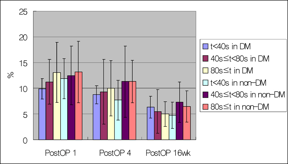
Figure 2. This shows the change of corneal endothelial cell density in postoperative periods in diabetics and non-diabetics (%). The changes were not statistically significantly different 1 week, 4 weeks, and 16 weeks after cataract surgery both in diabetics and in non-diabetics (P>0.05).
Reference
-
References
1. Olson RJ, Mamalis N, Werner L, Apple DJ. Cataract treatment in the beginning of the 21st century. Am J Ophthalmol. 2003; 136:146–54.
Article2. Lundstrom M, Stenevi U, Thorburn W. The Swedish National Cataract Register: A 9-year review. Acta Ophthalmol Scand. 2002; 80:248–57.3. Edwards M, Rehman S, Hood A, et al. Discharging routine phacoemulsification patients at one week. Eye. 1997; 11:850–3.
Article4. Bourne RR, Minassian DC, Dart JK, et al. Effect of cataract surgery on the corneal endothelium: modern phacoemulsification compared with extracapsular cataract surgery. Ophthalmology. 2004; 111:679–85.5. Binder PS, Sternberg H, Wickhan MG, Worthen DM. Corneal endothelial damage associated with phacoemulsification. Am J Ophthalmol. 1976; 82:48–54.6. Olson LE, Marshall J, Rice NS, Andrews R. Effects of ultrasound on the corneal endothelium: I. The acute lesion. Br J Ophthalmol. 1978; 62:134–44.
Article7. Walkow T, Anders N, Klebe S. Endothelial cell loss after phacoemulsification: Relation to preoperative parameters. J Cataract Refract Surg. 2000; 26:727–32.8. Larsson L-I, Bourne WM, Pach JM, Brubaker RF. Structure and function of the corneal endothelium in diabetes mellitus type I and II. Arch Ophthalmol. 1996; 114:9–14.9. Whikehart DR, Montgomery B, Angelos P, Sorna D. Alteration of ATPase activity and duplex DNA in corneal cells grown in high glucose media. Cornea. 1993; 12:295–8.
Article10. Schultz RO, Matsuda M, Yee RW, et al. Corneal endothelial changes in type I and type II diabetic mellitus. Am J Ophthalmol. 1984; 98:401–10.11. Itoi M, Nakamura T, Mizobe K, et al. Specular microscopic studies of the corneal endothelia of Japanese diabetics. Cornea. 1989; 8:2–6.
Article12. Straatsma BR, Pettit TH, Wheeler N, Miyamasu W. Diabetes mellitus and intraocular lens implantation. Ophthalmology. 1983; 90:336–43.
Article13. Lass JH, Spurney RV, Dutt RM, et al. A morphologic and fluorophotometric analysis of the corneal endothelium in type I diabetes mellitus and cystic fibrosis. AM J Ophthalmol. 1985; 100:783–8.
Article14. Laule A, Cable MK, Hoffman CE, Hanna C. Endothelial cell population changes of human cornea during life. Arch Ophthalmol. 1978; 96:2031–5.
Article15. Gilbert Smolin, Richard A, Thoft . The Cornea. 3rd ed. 1. Boston: Williams & Wilkins;1993. p. 25–42.16. Sugar J, Mitchelson J, Kraff M. The effect of phacoemulsification on corneal endothelial cell density. Arch Ophthalmol. 1978; 96:446–8.
Article17. Matsuda M, Miyake K, Inaba M. Long-term corneal endothelial changes after intraocular lens implantation. Am J Ophthalmol. 1988; 105:248–52.
Article18. Masket S. One year postop. Astigmatic comparison of sutured and unsutured from scleral pocket incision. J Cataract Refract Surg. 1993; 19:453–6.19. Gobbels M, Spitznas M. Endothelial barrier function after phacoemulsification: a comparison between diabetic and non-diabetic patients. Graefes Arch Clin Exp Ophthalmol. 1991; 229:254–7.20. Kosrirukvongs P, Slade S, Berkeley RG. Corneal endothelial changes after divide and conquer versus chip and flip phacoemulsification. J Cataract Refract Surg. 1997; 23:1006–12.
Article21. Altintas AG, Yilmaz E, Anavol MA, Can I. Comparison of corneal edema caused by cataract surgery with different phaco times in diabetic and non-diabetic patients. Ann Ophalmol. 2006; 38:61–5.
Article22. Shetlar DJ, Bourne WM, Campbell RJ. Morphologic evaluation of Descemet's membrane and corneal endothelium in diabetes mellitus. Ophthalmology. 1989; 96:247–50.
Article23. Lass JH, Spurney RV, Dutt RM, et al. A morphologic and fluorophotometric analysis of the corneal endothelium in type I diabetes mellitus and cystic fibrosis. Am J Ophthalmol. 1985; 100:783–8.
Article24. Zetterstrom C, Laurell CG. Comparison of endothelial cell loss and phacoemulsification energy during endocapsular phacoemulsification surgery. J Cataract Refract Surg. 1995; 21:55–8.25. Furuse N, Hayasaka S, Yamamoto Y, Setogawa T. Corneal endothelial changes after posterior chamber intraocular lens implantation in patients with or without diabetes mellitus. Br J Ophthalmol. 1990; 74:258–60.
Article26. Revalico G, Tognetto D, Palomba MA, et al. Corneal endothelial function after extracapsular cataract extraction and phacoemulsification. J Cataract Refract Surg. 1997; 23:1000–9.27. Yee RW, Matsuda M, Kern TS, et al. Corneal endothelial changes in diabetic dogs. Curr Eye Res. 1985; 4:759–66.
Article28. Dick HB, Kohnen T, Jacobi FK, Jacobi KW. Long-term endothelial cell loss following phacoemulsification through a temporal clear corneal incision. J Cataract Refract Surg. 1996; 22:63–71.
Article
- Full Text Links
-
- Actions
-
Cited
- CITED
-
- Close
- Share
-
- Similar articles
-
- The Effects of PhacoemulsificationTime and BSS Volume during Phacoemulsification on the Postoperative Corneal Endothelial Cell Loss
- The Effect of Phacoemulsification on the Corneal Endothelium in Diabetic Patients
- Corneal Endothelial Cell Change after Phacoemulsification according to the Severity of Diabetic Retinopathy
- The Effect of Phacoemulsification with a Liquefaction Device on Cornea Endothelium
- Corneal Endothelial Change after Clear Corneal Incision and Scleral Pocket Incision
