J Korean Ophthalmol Soc.
2007 Nov;48(11):1459-1465.
Cosmetic Repair of Band Keratopathy Using Stained Amniotic Membrane
- Affiliations
-
- 1Department of Ophthalmology, Seoul National University College of Medicine, Korea.
- 2Seoul Artificial Eye Center, Seoul National University Hospital Clinical Research Institute, Seoul, Korea. eyeminerva@yahoo.co.kr
- 3Department of Ophthalmology, Seoul National University Boramae Hospital, Seoul, Korea.
- 4Department of Ophthalmology, Seoul National University Bundang Hospital, Gyeonggi, Korea.
- 5Seoul National University Hospital Healthcare System Gangnam Center, Healthcare Institute, Seoul, Korea.
Abstract
-
PURPOSE: To evaluate the efficacy of stained amniotic membrane transplantation (AMT) for cosmetic repair of band keratopathy.
METHODS
Eight eyes from 8 patients with band keratopathy without the possibility of visual improvement were treated with corneal tattooing and stained AMT. Calcific plaques were removed with EDTA and a blade prior to the tattooing. Tattooing by the injection of tissue marking dye into the corneal stroma was performed in corneal opacities. The remaining opacity, which could not be treated with corneal tattooing, was treated with black dye-stained AMT.
RESULTS
The most common cause of band keratopathy was trauma (50%) and the mean time to epithelial healing was 11.3 days. All patients showed satisfactory results with cosmetic staining. Recurring cases and significant complications were not observed during the mean follow-up period of 7.4 months.
CONCLUSIONS
Cosmetic repair by corneal tattooing and stained AMT is an effective treatment for band keratopathy and is not associated with any significant complications.
MeSH Terms
Figure
-
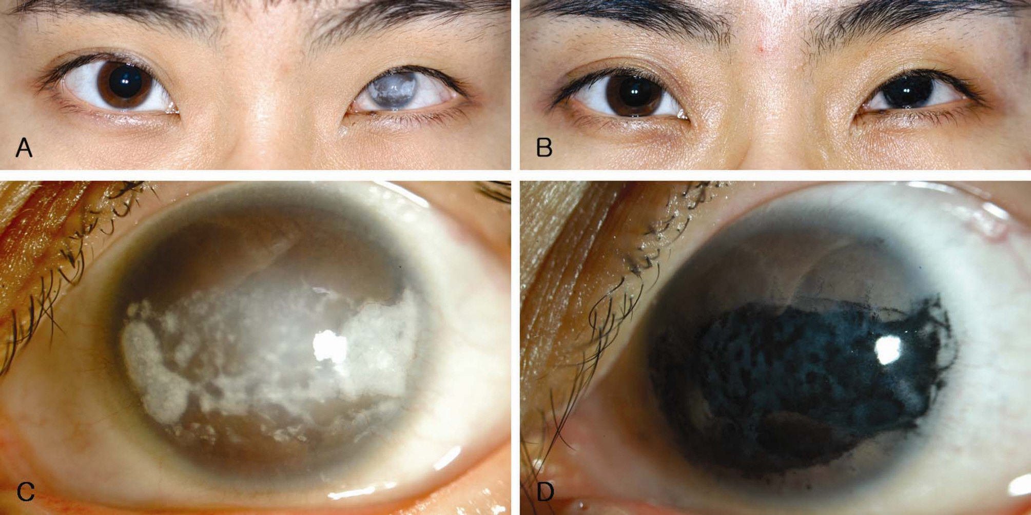
Figure 1. A 20-year-old female patient with band keratopathy in the left eye. (A) and (C): Before tattooing and stained amniotic membrane transplantation (AMT). (B) and (D): After tattooing and stained AMT.
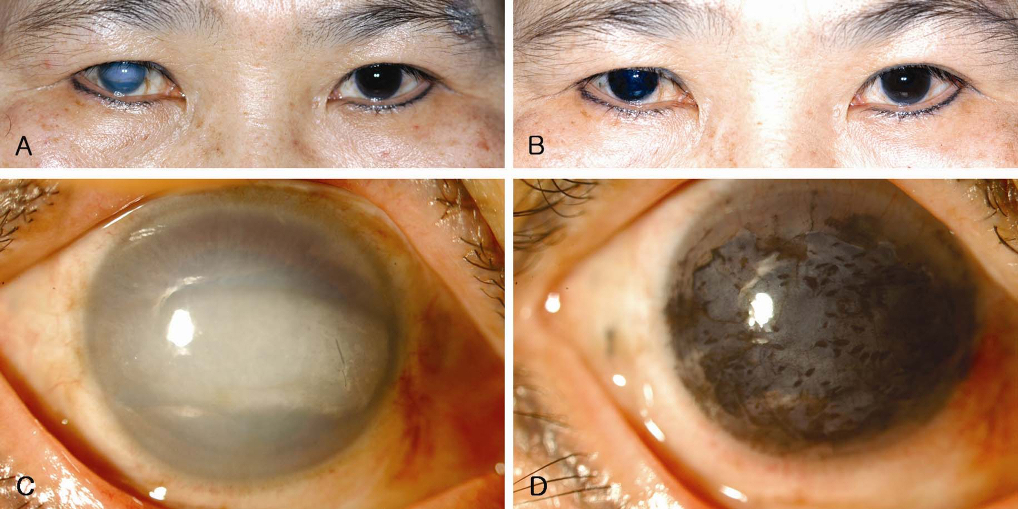
Figure 2. A 63-year-old female patient with band keratopathy in the right eye. (A) and (C): Before tattooing and stained AMT. (B) and (D): After tattooing and stained AMT.
Reference
-
References
1. Brazier DJ, Hitchings RA. Atypical band keratopathy following long-term pilocarpine treatment. Br J Ophthalmol. 1989; 73:294–6.
Article2. Najjar DM, Cohen EJ, Rapuano CJ, Laibson PR. EDTA chelation for calcific band keratopathy: results and long-term follow-up. Am J Ophthalmol. 2004; 137:1056–64.
Article3. Bokosky JE, Meyer RF, Sugar A. Surgical treatment of calcific band keratopathy. Ophthalmic Surg. 1985; 16:645–7.
Article4. Baltatzis S, Papaefthimiou J. Treatment of calcific band keratopathy by Nd:YAG laser. Eur J Ophthalmol. 1992; 2:27–9.5. O’ Brart DPS, Gartry DS, Lohmann CP, et al. Treatment of band keratopathy by excimer laser phototherapeutic keratectomy: surgical techniques and long term follow up. Br J Ophthalmol. 1993; 77:702–8.6. Dighiero P, Boudraa R, Ellies P, et al. Therapeutic photokeratectomy for the treatment of band keratopathy. Jr F Ophthalmol. 2000; 23:345–9.7. Stewart OG, Morrell AJ. Management of band keratopathy with excimer phototherapeutic keratectomy: visual, refractive, and symptomatic outcome. Eye. 2003; 17:233–7.
Article8. Anderson DF, Prabhasawat P, Alfonso E, Tseng SCG. Amniotic membrane transplantation after the primary surgical management of band keratopathy. Cornea. 2001; 20:354–61.
Article9. Kwon YS, Song YS, Kim JC. New treatment for band keratopathy: superficial lamellar keratectomy, EDTA chelation and amniotic membrane transplantation. J Korean Med Sci. 2004; 19:611–5.
Article10. Kim C, Han YK, Wee WR, et al. Cosmetic repair of corneal opacity by tattooing. J Korean Ophthalmol Soc. 2005; 46:1967–73.11. Arjamaa O. EDTA chelation for calcific band keratopathy. Am J Ophthalmol. 2005; 139:216.
Article12. Cursino JW, Fine BS. A histologic study of calcific and noncalcific band keratopathies. Am J Ophthalmol. 1976; 82:395–404.
Article13. Meller D, Pires RT, Tseng SC. Ex vivo preservation and expansion of human limbal epithelial stem cells on amniotic membrane cultures. Br J Ophthalmol. 2002; 86:463–71.
Article14. Tseng SC, Prabhasawat P, Lee SH. Amniotic membrane transplantation for conjunctival surface reconstruction. Am J Ophthalmol. 1997; 124:765–74.
Article15. Tseng SC, Prabhasawat P, Barton K, et al. Amniotic membrane transplantation with or without limbal allografts for corneal surface reconstruction in patients with limbal stem cell deficiency. Arch Ophthalmol. 1998; 116:431–41.
Article16. Kim JC, Tseng SC. Transplantation of preserved human amniotic membrane for surface reconstruction in severely damaged rabbit corneas. Cornea. 1995; 14:473–84.
Article17. Gomes JA, Romano A, Santos MS, Dua HS. Amniotic membrane use in ophthalmology. Curr Opin Ophthalmol. 2005; 16:233–40.
Article18. Kenyon KR. Amniotic membrane: mother's own remedy for ocular surface disease. Cornea. 2005; 24:639–42.19. Lee JE, Jun JB, Choi HY, et al. Corneal tattooing to mask subsequent opacification after amniotic membrane grafting for stromal corneal ulcer. Acta Ophthalmol Scand. 2006; 84:696–8.
Article20. Tang CK, Ahn HB, Park WC. The clinical effects of dye-amniotic membrane transplantation on bullous keratopathy. J Korean Ophthalmol Soc. 2003; 44:1741–7.
- Full Text Links
-
- Actions
-
Cited
- CITED
-
- Close
- Share
-
- Similar articles
-
- The Clinical Effects of Dye-Amniotic Membrane Transplantation on Bullous Keratopathy
- New Treatment for Band Keratopathy: Superficial Lamellar Keratectomy, EDTA Chelation and Amniotic Membrane Transplantation
- 7 Cases of Combined Corneal Tattooing and Amniotic Membrane Transplantation in Bullous Keratopathy
- Combined Ethylenediaminetetraacetic Acid Chelation, Phototherapeutic Keratectomy and Amniotic Membrane Transplantation for Treatment of Band Keratopathy
- Combined Phototherapeutic Keratectomy and Amniotic Membrane Transplantation in the Treatment of Bullous Keratopathy
