J Korean Ophthalmol Soc.
2007 Aug;48(8):1158-1162.
Two Cases of Ocular Ischemia following Scleral Encircling
- Affiliations
-
- 1Kong Eye Center, Seoul, Korea. euklee@chol.com
Abstract
-
PURPOSE: To report two cases of ocular ischemia following scleral encircling.
METHODS
A 21-year-old man with glaucoma and a 76-year-old woman without any medical problem were transferred to our department for surgery to treat retinal detachment. After retrobulbar anesthesia and limbal peritomy of conjunctiva, the 4-rectus muscles were isolated. Scleral encircling was performed with No. a 42 band (4.0 mm in width) after cryotherapy done completely around retinal tear.
RESULTS
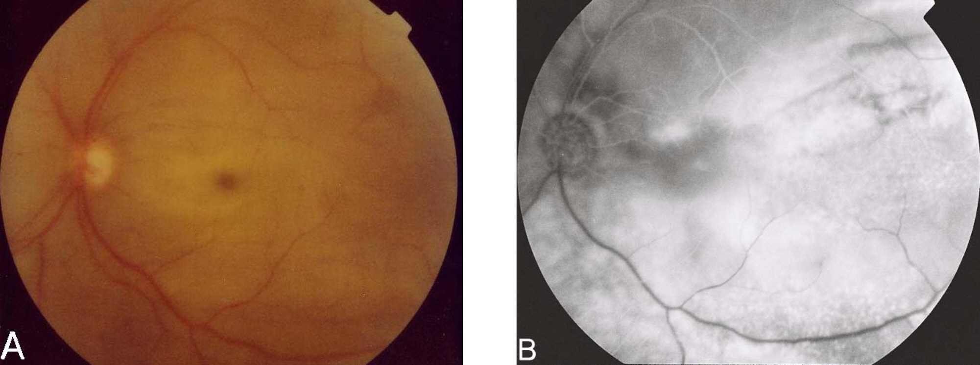
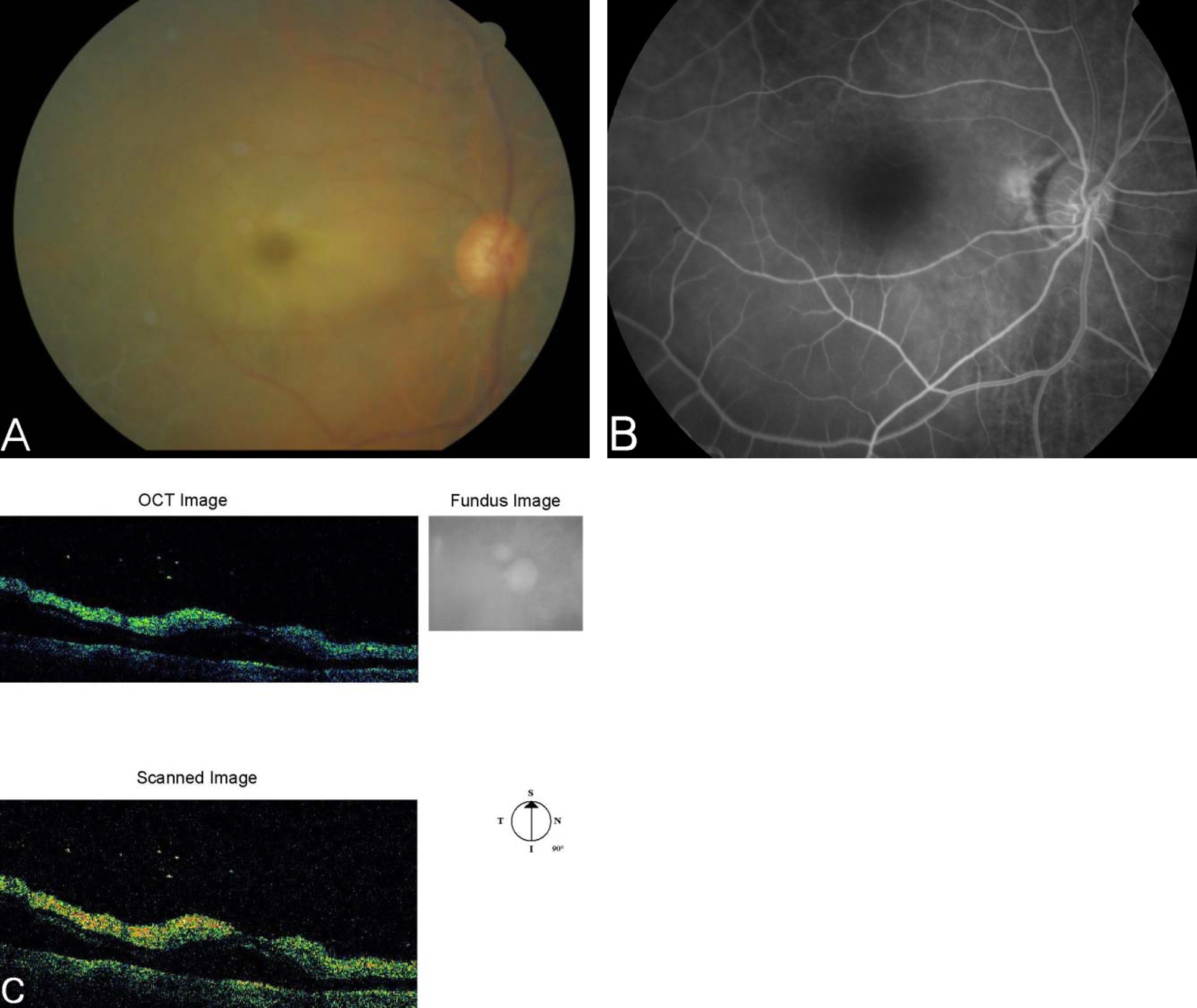
Following surgery, One patient experienced ophthalmic artery occlusion and while the other patient experienced central retinal artery occlusion. Vision was not restored in either cases despite IV injection of 250 ml of 15% mannitol solution and anterior chamber paracentesis.
CONCLUSIONS
In the cases where patients are of old age or suffer from glaucoma, we strongly recommend that the surgeons perform the scleral encircling carefully.
MeSH Terms
Figure
Reference
-
References
1. Schepens CL. Management of retinal detachment. Ophthalmic Surg. 1994; 25:727–31.
Article2. Gonin J. The treatment of detached retina by searing the retinal tears. Arch Ophthalmol. 1930; 4:621–5.
Article3. Lincoff HA, Baros I, McLean J. Modification to the Custodis procedure for retinal detachment. Arch Ophthalmol. 1965; 73:160–3.4. Schepens CL. Symposium: Present status of retinal detachment surgery-Scleral buckling with circling element. Trans Am Acad Ophthalmol Otolaryngol. 1964; 68:959–79.5. Edmund J, Seedorff HH. Retinal detachment surgery II. Acta Ophthalmol. 1968; 46:643–65.6. Greven CM, Wall AB, Slusher MM. Anatomic and visual results in asymptomatic clinical rhegmatogenous retinal detachment repaired by scleral buckling. Am J Ophthalmol. 1999; 128:618–20.
Article7. Yi Yao, Li Jiang, Zhi-jun wang, Mao-nian Zhang. Scleral buckling procedures for longstanding or chronic rhegmatogenous retinal detachment with subretinal proliferation. Ophthalmology. 2006; 113:821–5.
Article8. Han DP, Mohsin NC, Guse CE. Comparision of Pneumatic retinopexy and scleral buckling in the management of primary rhegmatogenous retinal detachment. Am J Ophthalmol. 1998; 126:658–68.9. Lee MV, Moon CS, Yang HS, et al. Factor influencing anatomic failure of simple rhegmatogenous retinal detachment. J Korean Ophthalmol Soc. 2006; 47:407–14.10. Ahmadieh H, Entezari M, Soheilian M. Factors influencing anatomic and visual results primary scleral buckling. Eur J Ophthalmol. 2000; 10:153–9.11. Burton TC. Preoperative factors influencing anatomic success rates following retinal detachment surgery. Trans Am Acad Ophthalmol Otolaryngol. 1997; 83:499–505.12. Park JL, Kim SD, Yun IH. A clinical study of the rhegmatogenous retinal detachment. J Korean Ophthalmol Soc. 2002; 43:1015–24.13. Laatikaien L, Toippanen EM. Characteristics of rhegmatogenous retinal detachment. Acta Ophthalmol. 1985; 63:146–54.14. Laatikaien L, Harju H, Toippanen EM. Postoperative outcome in rhegmatogenous retinal detachment. Acta Ophthalmol. 1985; 63:647–55.15. Burton TC, Lambert RW. A predictive model for vision recovery following retinal detachment surgery. Ophthamology. 1978; 85:619–25.16. Kreiger AE, Hodgkinson BJ, Fredrick AR, et al. The results of retinal detachment surgery-Analysis of 268 operations with briad scleral buckling. Arch Ophthalmol. 1971; 86:385–94.17. Park JI, Shim WS. A clinical study of the retinal detachment surgery utilizing silicone rubber. J Korean Ophthalmol Soc. 1980; 21:435–40.18. Park BK, Jo JI. A clinical study of the retinal detachment. J Korean Ophthalmol Soc. 1981; 22:787–97.19. Jeong SK, Park YG, Lee MK. A clinical study on rhegmatogenous retinal detachment. J Korean Ophthalmol Soc. 1992; 33:589–98.20. Jo SM, Kim SY, Kwoon OW. Effect of encircling sponge for retinal detachment. J Korean Ophthalmol Soc. 1992; 33:1070–6.21. Hayreh SS, Zimmerman MB, Kimura A, et al. Central retinal artery occlusion-Retinal survival time. Exp Eye Res. 2004; 78:723–6.
- Full Text Links
- Actions
-
Cited
- CITED
-
- Close
- Share
- Similar articles
-
- Effect of IOP elevation on matrix metalloproteinase-2 in rabbit anterior chamber
- Recurrence of Retinal Detachment after Scleral Buckle Removal
- Characteristics of Scleral Lenses and Patient Selection
- Pefractive Changes After Encircling Scleral Buckling Surgery
- 7 Cases of Cryopexy and Full-Thickness Scleral Buckling with Encircling Band in Retinal Detachment


