Obstet Gynecol Sci.
2016 Jul;59(4):303-306. 10.5468/ogs.2016.59.4.303.
Reference values for the cervical length measurement in the second trimester of pregnancy using the transvaginal ultrasound in a large Brazilian population
- Affiliations
-
- 1Mario Palmério University Hospital, University of Uberaba, Uberaba-MG, Brazil.
- 2Radiologic Clinic of Uberaba, Uberaba-MG, Brazil.
- 3Department of Obstetrics and Gynecology, Ribeirão Preto Medical School, University of São Paulo, Ribeirão Preto-SP, Brazil.
- 4Department of Obstetrics and Gynecology, Federal University of Paraná, Curitiba-PR, Brazil.
- 5Department of Obstetrics, Paulista School of Medicine, Federal University of São Paulo, São Paulo-SP, Brazil. araujojred@terra.com.br
- KMID: 2329051
- DOI: http://doi.org/10.5468/ogs.2016.59.4.303
Abstract
- To establish reference values for the cervical length (CL) measurement by transvaginal ultrasound between 20 and 24+6 weeks of gestation in a large Brazilian population. A retrospective cross-sectional study was performed with 996 singleton pregnancies. The CL measurement (mm) using the transvaginal ultrasound was obtained in a sagittal view and the calipers positioned to measure the linear distance between the triangular area of echodensity at the external os and the internal os. The median±standard deviation and ranges for the CL measurement (mm) was 37.0±10.7 (range, 8 to 51). CL measurement did not modify significantly with gestational age. The observed percentiles for the CL measurement (mm) considering all number case were the following: 5th, 28 mm; 50th, 37 mm; and 95th, 45 mm. Reference values for the CL measurement by transvaginal ultrasound between 20 and 24+6 weeks of gestation in a large heterogeneous Brazilian population were established.
MeSH Terms
Figure
-
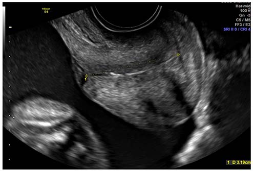
Fig. 1 Transvaginal ultrasound examination showing the cervical length measurement at 22 weeks of gestation.
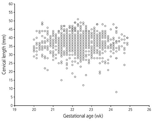
Fig. 2 Scatterplot for cervical length measurement (mm) as a function of gestational age (weeks).
Reference
-
1. Guimaraes Filho HA, Araujo Junior E, Pires CR, Nardozza LM, Moron AF. Short cervix syndrome: current knowledge from etiology to the control. Arch Gynecol Obstet. 2013; 287:621–628.2. Martin JA, Hamilton BE, Sutton PD, Ventura SJ, Mathews TJ, Osterman MJ. Births: final data for 2008. Natl Vital Stat Rep. 2010; 59:13–71.3. Goldenberg RL, Culhane JF, Iams JD, Romero R. Epidemiology and causes of preterm birth. Lancet. 2008; 371:75–84.4. Haram K, Mortensen JH, Wollen AL. Preterm delivery: an overview. Acta Obstet Gynecol Scand. 2003; 82:687–704.5. Andersen HF, Nugent CE, Wanty SD, Hayashi RH. Prediction of risk for preterm delivery by ultrasonographic measurement of cervical length. Am J Obstet Gynecol. 1990; 163:859–867.6. Fonseca EB, Celik E, Parra M, Singh M, Nicolaides KH. Fetal Medicine Foundation Second Trimester Screening Group. Progesterone and the risk of preterm birth among women with a short cervix. N Engl J Med. 2007; 357:462–469.7. Cahill AG, Odibo AO, Caughey AB, Stamilio DM, Hassan SS, Macones GA, et al. Universal cervical length screening and treatment with vaginal progesterone to prevent preterm birth: a decision and economic analysis. Am J Obstet Gynecol. 2010; 202:548.e1–548.e8.8. Stein W, Hellmeyer L, Schmidt S, Tekesin I. Intraobserver and interobserver reliability of transvaginal cervical length measurements and quantitative ultrasound tissue characterization of the cervix in the second and third trimester of pregnancy. Ultraschall Med. 2011; 32:Suppl 2. E169–E174.9. Hibbard JU, Tart M, Moawad AH. Cervical length at 16-22 weeks' gestation and risk for preterm delivery. Obstet Gynecol. 2000; 96:972–978.10. Rovas L, Sladkevicius P, Strobel E, Valentin L. Reference data representative of normal findings at two-dimensional and three-dimensional gray-scale ultrasound examination of the cervix from 17 to 41 weeks' gestation. Ultrasound Obstet Gynecol. 2006; 27:392–402.11. Erasmus I, Nicolaou E, van Gelderen CJ, Nicolaides KH. Cervical length at 23 weeks' gestation: relation to demographic characteristics and previous obstetric history in South African women. S Afr Med J. 2005; 95:691–695.12. Silva SV, Damiao R, Fonseca EB, Garcia S, Lippi UG. Reference ranges for cervical length by transvaginal scan in singleton pregnancies. J Matern Fetal Neonatal Med. 2010; 23:379–382.13. Sonek J, Shellhaas C. Cervical sonography: a review. Ultrasound Obstet Gynecol. 1998; 11:71–78.14. Altman DG, Chitty LS. Charts of fetal size: 1. Methodology. Br J Obstet Gynaecol. 1994; 101:29–34.15. Barros-Silva J, Pedrosa AC, Matias A. Sonographic measurement of cervical length as a predictor of preterm delivery: a systematic review. J Perinat Med. 2014; 42:281–293.16. Alfirevic Z, Owen J, Carreras Moratonas E, Sharp AN, Szychowski JM, Goya M. Vaginal progesterone, cerclage or cervical pessary for preventing preterm birth in asymptomatic singleton pregnant women with a history of preterm birth and a sonographic short cervix. Ultrasound Obstet Gynecol. 2013; 41:146–151.17. Instituto Brasileiro de Geografia e Estatistica. Miscegenation decreases percentage of black and white [Internet]. [place unknown]: Instituto Brasileiro de Geografia e Estatistica;cited 2007 May 25. Available from: http://www.ibge.com.br/home/presidencia/noticias/noticia_visualiza.php?id_noticia=892&id_pagina=1.
- Full Text Links
-
- Actions
-
Cited
- CITED
-
- Close
- Share
-
- Similar articles
-
- Cervical Length During Pregnancy in Korean Women as Assessed by Transvaginal Ultrasound: A Prospective Longitudinal Study
- Normal Reference Range for Cervical Length in Twin Pregnancy using transvaginal ultrasound
- Role of ultrasound in the evaluation of first-trimester pregnancies in the acute setting
- Prediction of Prolonged Pregnancy in Nulliparous Women by Transvaginal Ultrasonographic Measurement of Cervical Length at 20-24 Weeks and 37 Weeks
- Nuchal Translucency Measurement in Normal Korean Fetuses at 10-14 Weeks of Gestation(II)
