Occult Intertrochanteric Fracture Mimicking the Fracture of Greater Trochanter
- Affiliations
-
- 1Department of Orthopedic Surgery, Dongguk University College of Medicine, Gyeongju, Korea. shoulder2011@dongguk.ac.kr
- KMID: 2328347
- DOI: http://doi.org/10.5371/hp.2016.28.2.112
Abstract
- PURPOSE
Occult intertrochanteric fractures are misdiagnosed as isolated greater trochanteric fractures in some cases. We investigated the utility of three-dimensional computed tomography (3D-CT) and magnetic resonance imaging (MRI) in the diagnosis and outcome management of occult intertrochanteric fractures.
MATERIALS AND METHODS
This study involved 23 cases of greater trochanteric fractures as diagnosed using plain radiographs from January 2004 to July 2013. Until January 2008, 9 cases were examined with 3D-CT only, while 14 cases were screened with both 3D-CT and MRI scans. We analyzed diagnostic accuracy and treatment results following 3D-CT and MRI scanning.
RESULTS
Nine cases that underwent 3D-CT only were diagnosed with isolated greater trochanteric fractures without occult intertrochanteric fractures. Of these, a patient with displacement received surgical treatment. Of the 14 patients screened using both CT and MRI, 13 were diagnosed with occult intertrochanteric fractures. Of these, 11 were treated with surgical intervention and 2 with conservative management.
CONCLUSION
Three-dimensional CT has very low diagnostic accuracy in diagnosing occult intertrochanteric fractures. For this reason, MRI is recommended to confirm a suspected occult intertrochanteric fracture and to determine the most appropriate mode of treatment.
Keyword
Figure
-
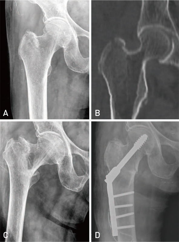
Fig. 1 A 91-year-old woman who presented with right hip pain after fall down. (A) Initial radiograph shows isolated greater trochanteric fracture. (B) Three-dimensional computed tomography shows no occult intertrochanteric fracture. (C) At 9 days after trauma, the radiography shows a displaced intertrochanteric fracture. (D) The postoperative radiograph shows fixation with compressive hip screw.
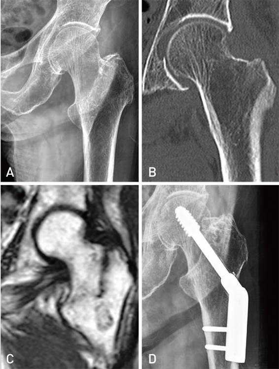
Fig. 2 A 68-years-old woman who presented with left hip pain after fall down. (A) Initial radiograph shows isolated greater trochanteric fracture. (B) Three-dimensional computed tomography shows no occult intertrochanteric fracture. (C) Coronal T1-weighted MRI revealed a fracture from the greater trochanter leading toward the lesser trochanter. (D) The postoperative radiograph shows fixation with compressive hip screw.
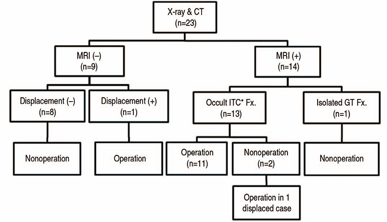
Fig. 3 Summary of diagnosis and treatment. CT: computed tomography, MRI: magnetic resonance imaging, ITC: intertrochanter, Fx.: fracture, GT: greater trochanter.
Cited by 1 articles
-
The Frequency of Occult Intertrochanteric Fractures among Individuals with Isolated Greater Trochanteric Fractures
Jongho Noh, Kee Haeng Lee, Sehoon Jung, Sunwook Hwang
Hip Pelvis. 2019;31(1):23-32. doi: 10.5371/hp.2019.31.1.23.
Reference
-
1. Chatha H, Ullah S, Cheema Z. Review article: Magnetic resonance imaging and computed tomography in the diagnosis of occult proximal femur fractures. J Orthop Surg (Hong Kong). 2011; 19:99–103.
Article2. Iwata T, Nozawa S, Dohjima T, et al. The value of T1-weighted coronal MRI scans in diagnosing occult fracture of the hip. J Bone Joint Surg Br. 2012; 94:969–973.
Article3. Reiter M, O'Brien SD, Bui-Mansfield LT, Alderete J. Greater trochanteric fracture with occult intertrochanteric extension. Emerg Radiol. 2013; 20:469–472.
Article4. Lee KH, Kim HM, Kim YS, et al. Isolated fractures of the greater trochanter with occult intertrochanteric extension. Arch Orthop Trauma Surg. 2010; 130:1275–1280.
Article5. Holder LE, Schwarz C, Wernicke PG, Michael RH. Radionuclide bone imaging in the early detection of fractures of the proximal femur (hip): multifactorial analysis. Radiology. 1990; 174:509–515.
Article6. Omura T, Takahashi M, Koide Y, et al. Evaluation of isolated fractures of the greater trochanter with magnetic resonance imaging. Arch Orthop Trauma Surg. 2000; 120:195–197.
Article7. Craig JG, Moed BR, Eyler WR, van Holsbeeck M. Fractures of the greater trochanter: intertrochanteric extension shown by MR imaging. Skeletal Radiol. 2000; 29:572–576.
Article8. Feldman F, Staron RB. MRI of seemingly isolated greater trochanteric fractures. AJR Am J Roentgenol. 2004; 183:323–329.
Article9. Frihagen F, Nordsletten L, Tariq R, Madsen JE. MRI diagnosis of occult hip fractures. Acta Orthop. 2005; 76:524–530.
Article10. Beloosesky Y, Hershkovitz A, Guz A, Golan H, Salai M, Weiss A. Clinical characteristics and long-term mortality of occult hip fracture elderly patients. Injury. 2010; 41:343–347.
Article11. Koval KJ, Skovron ML, Aharonoff GB, Meadows SE, Zuckerman JD. Ambulatory ability after hip fracture. A prospective study in geriatric patients. Clin Orthop Relat Res. 1995; (310):150–159.12. Koval KJ, Zuckerman JD. Functional recovery after fracture of the hip. J Bone Joint Surg Am. 1994; 76:751–758.
Article13. Kanis JA, Johnell O, De Laet C, Jonsson B, Oden A, Ogelsby AK. International variations in hip fracture probabilities: implications for risk assessment. J Bone Miner Res. 2002; 17:1237–1244.
Article14. Gullberg B, Johnell O, Kanis JA. World-wide projections for hip fracture. Osteoporos Int. 1997; 7:407–413.
Article15. Rizzo PF, Gould ES, Lyden JP, Asnis SE. Diagnosis of occult fractures about the hip. Magnetic resonance imaging compared with bone-scanning. J Bone Joint Surg Am. 1993; 75:395–401.
Article16. Feldman F, Staron R, Zwass A, Rubin S, Haramati N. MR imaging: its role in detecting occult fractures. Skeletal Radiol. 1994; 23:439–444.
Article17. Cabarrus MC, Ambekar A, Lu Y, Link TM. MRI and CT of insufficiency fractures of the pelvis and the proximal femur. AJR Am J Roentgenol. 2008; 191:995–1001.
Article18. Matin P. The appearance of bone scans following fractures, including immediate and long-term studies. J Nucl Med. 1979; 20:1227–1231.19. Ingari JV, Smith DK, Aufdemorte TB, Yaszemski MJ. Anatomic significance of magnetic resonance imaging findings in hip fracture. Clin Orthop Relat Res. 1996; (332):209–214.
Article20. Quinn SF, McCarthy JL. Prospective evaluation of patients with suspected hip fracture and indeterminate radiographs: use of T1-weighted MR images. Radiology. 1993; 187:469–471.
Article21. Alam A, Willett K, Ostlere S. The MRI diagnosis and management of incomplete intertrochanteric fractures of the femur. J Bone Joint Surg Br. 2005; 87:1253–1255.
Article22. Rubin G, Malka I, Rozen N. Should we operate on occult hip fractures? Isr Med Assoc J. 2010; 12:316–317.23. Schultz E, Miller TT, Boruchov SD, Schmell EB, Toledano B. Incomplete intertrochanteric fractures: imaging features and clinical management. Radiology. 1999; 211:237–240.
Article
- Full Text Links
-
- Actions
-
Cited
- CITED
-
- Close
- Share
-
- Similar articles
-
- Delayed Pseudoaneurysm of Deep Femoral Artery Caused by Migration of Lesser Trochanter, Subsequent to an Intertrochanteric Fracture Surgery - A Case Report -
- Bipolar Hemiarthroplasty Using the Greater Trochanter Reattachment Device (GTRD) for Comminuted Intertrochanteric Femur Fracture in Elderly Patients
- Changes of the Fracture Fragments of Lesser Trochanter after Operative Treatment in the Unstable Femoral Intertrochanteric Fractures
- Modified Double Tension Band Wiring for Reattaching the Greater Trochanter When Performing Hemiarthroplasty for Intertrochanteric Fracture in Elderly Patients
- New Wiring Method for Lesser Trochanter Fixation in Unstable Intertrochanteric Fractures: Technical Note
