Investig Magn Reson Imaging.
2016 Jun;20(2):136-139. 10.13104/imri.2016.20.2.136.
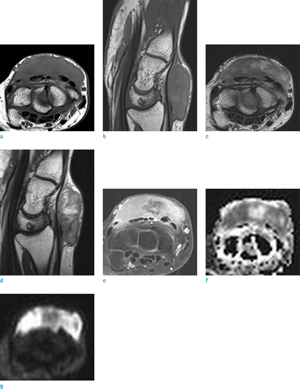
Clear Cell Sarcoma of the Wrist: MRI Findings with Diffusion-Weighted Image and Histopathologic Correlation
- Affiliations
-
- 1Department of Radiology, Hallym University College of Medicine, Dongtan Sacred Heart Hospital, Gyeonggi-do, Korea. jachoi88@gmail.com
- 2Department of Pathology, Hallym University College of Medicine, Dongtan Sacred Heart Hospital, Gyeonggi-do, Korea.
- KMID: 2327428
- DOI: http://doi.org/10.13104/imri.2016.20.2.136
Abstract
- Clear cell sarcoma is rare and difficult to diagnose. Herein, we present a case of clear cell sarcoma in the dorsum of the wrist with MRI findings, including diffusion-weighted imaging, and histopathologic correlation, which was initially diagnosed as giant cell tumor of tendon sheath.
Keyword
MeSH Terms
Figure
Reference
-
1. Kransdorf MJ, Murphey M. Imaging of soft tissue tumors. 3rd ed. Philadelphia: Lippincott Williams & Wilkins;a WOLTERS KLUWER business;2014. p. 435–438.2. Hoffman GJ, Carter D. Clear cell sarcoma of tendons and aponeuroses with melanin. Arch Pathol. 1973; 95:22–25.3. De Beuckeleer LH, De Schepper AM, Vandevenne JE, et al. MR imaging of clear cell sarcoma (malignant melanoma of the soft parts): a multicenter correlative MRI-pathology study of 21 cases and literature review. Skeletal Radiol. 2000; 29:187–195.4. Maeda M, Matsumine A, Kato H, et al. Soft-tissue tumors evaluated by line-scan diffusion-weighted imaging: influence of myxoid matrix on the apparent diffusion coefficient. J Magn Reson Imaging. 2007; 25:1199–1204.5. Razek A, Nada N, Ghaniem M, Elkhamary S. Assessment of soft tissue tumours of the extremities with diffusion echoplanar MR imaging. Radiol Med. 2012; 117:96–101.6. Kindblom LG, Lodding P, Angervall L. Clear-cell sarcoma of tendons and aponeuroses. An immunohistochemical and electron microscopic analysis indicating neural crest origin. Virchows Arch A Pathol Anat Histopathol. 1983; 401:109–128.7. Panagopoulos I, Mertens F, Isaksson M, Mandahl N. Absence of mutations of the BRAF gene in malignant melanoma of soft parts (clear cell sarcoma of tendons and aponeuroses). Cancer Genet Cytogenet. 2005; 156:74–76.8. Park BM, Jin SA, Choi YD, et al. Two cases of clear cell sarcoma with different clinical and genetic features: cutaneous type with BRAF mutation and subcutaneous type with KIT mutation. Br J Dermatol. 2013; 169:1346–1352.
- Full Text Links
- Actions
-
Cited
- CITED
-
- Close
- Share
- Similar articles
-
- Clear Cell Sarcoma of Flexor Tendon in Wrist: A Case Report
- The Value of PROPELLER Diffusion-Weighted Image in the Detection of Cholesteatoma
- MRI Findings of Intracranial IVleningioma: Significance of Gd-DTPA Enhancernent
- A Case of Malignant Melanoma of Soft Parts with Unusual Histopathologic Findings
- The Usefulness of Diffusion: Weighted Magnetic Resonance Image in the Diagnosis of Neonatal Seizure


