J Korean Soc Spine Surg.
2002 Jun;9(2):127-132.
Radiologic Result of Displacement according to Position and Measurement Methods in Spondylolisthesis
- Affiliations
-
- 1Department of Orthopaedics Surgery, Wonkwang University Hospital, Iksan, Korea. oschae@hitel.net
Abstract
-
STUDY DESIGN: Prospective analysis was based on radiographic appearance in 80 cases of spondylolisthesis taken in positional change.
PURPOSE: The aim of the study was to investigate the flexion-extension lateral radiographs about the difference between decubitus and upright position and the measurement method of displacement in spondylolisthesis.
SUMMARY OF LITERATURE REVIEW: Although the flexion-extension lateral radiographs of spine were known the most preferable diagnostic method for spine instability, there are some debates about the difference of displacement according to the patient position and measurement methods.
MATERIALS AND METHODS
The radiographs of 80 patients with spondylolisthesis were taken in the decubitus and upright position. Extent of the displacement were measured by Taillard, DuPuis, modified Qunnell & Stockdale method and Ferguson angle, slip angle, lordosis angle and vertebral centroid measurement of lumbar lordosis(CLL) were measured, according to position.
RESULTS
Significant difference between the positions was shown on the CLL and lordosis angle. Differences between positions analyzed from Taillard, DuPuis, modified Qunnell & Stockdale method, Ferguson angle and slip angle had no statistical significance. Differences between positions analyzed from the pathologic movement of translation(>4 mm) had a clinically significance in the upright position rather than the decubitus.
CONCLUSION
The lateral flexion-extension radiographs on upright position rather than decubitus position are considered as the more useful diagnostic method.
Figure
-
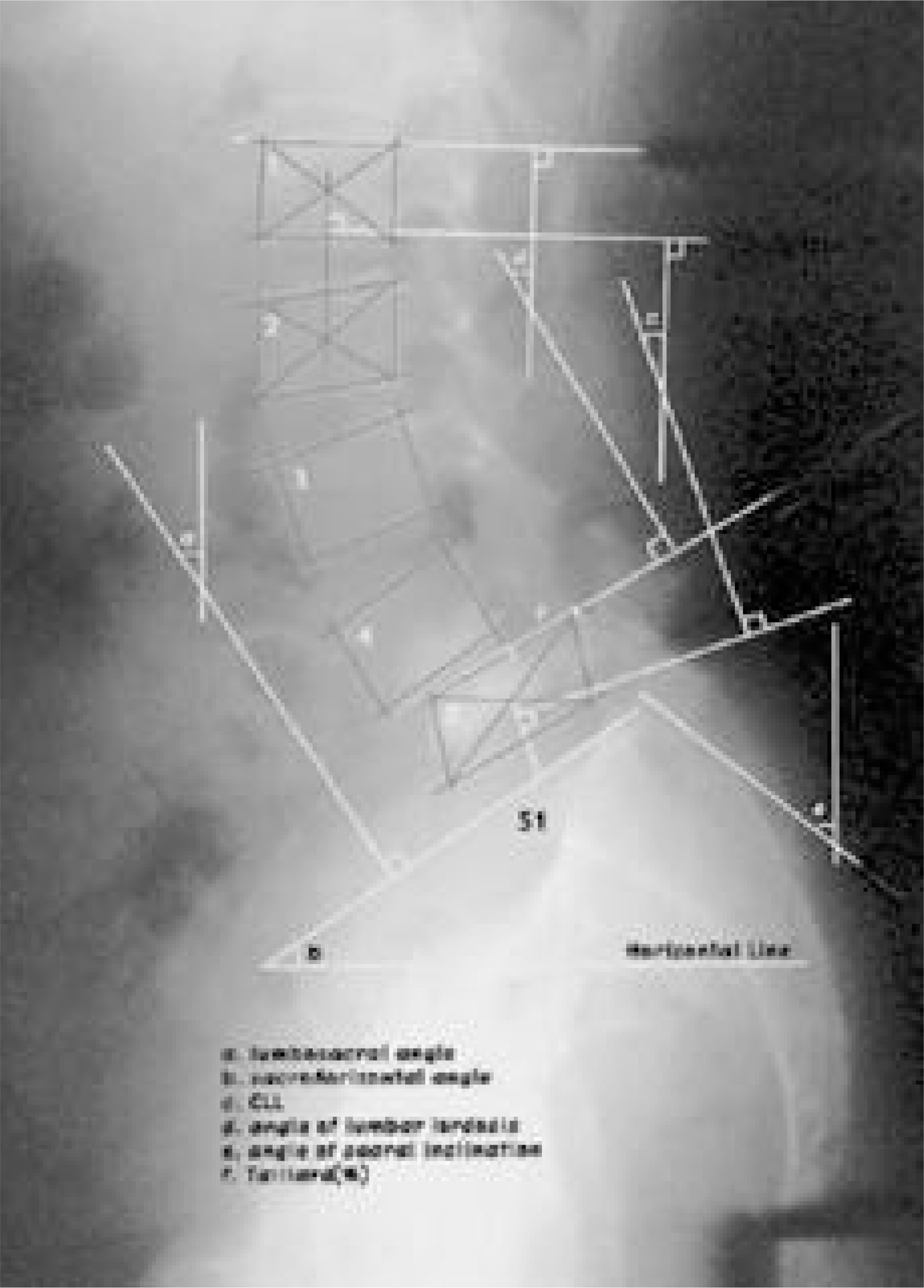
Fig. 1. Parameters that represents the radiographic measurement of the lumbar spine. a. lumbosacral angle b. sacro-horizontal angle c. CLL d. angle of lumbar lordosis e. angle of sacral inclination f. Taillard(%)
Reference
-
1). Bernhardt M, Bridwell KH. Segmental analysis of the sagittal plane alignment of the normal thoracic and lumbar spines and thoracolumbar junction. Spine. 14:717–721. 1989.
Article2). Chen YL. Vertebral centroid measurement of lumbar lordosis compared with the Cobb technique. Spine. 24:1786–1790. 1999.
Article3). Dupuis PR, Yong-hing K, Cassidy JD, Kirkaldy-Willis WH. Radiologic diagnosis of degenerative lumbar spinal instability. Spine. 10:262–276. 1985.
Article4). Farfan HF, Gracovetsky S. The nature of instability. Spine. 9:714–719. 1984.
Article5). Freiberg ORA. Lumbar instability: A dynamic approach by traction- compression radiography. Spine. 12:119–129. 1987.6). Frymoyer JW, Selby DK. Segmental instability. Spine. 10:280–287. 1985.
Article7). Inoue S, Watanabe T, Goto S, et al. D egenerative spondylolisthesis. Pathophysiology and results of anterior interbody fusion. Clin Orthop. 227:90–98. 1988.8). Junghanns H. Spondylolisthesis ohne. Spalt in Zwischen -gelenk-stuck. Arch Orthop Unfall Chir. 29:118–127. 1930.9). Knutsson F. The instability associated with disc degeneration in the lumbar spine. Acta Radiol. 25:593–609. 1944.10). Lowe RW, Hayes TD, Kaye J, Bagg RJ, Luekens CA. Standing roentgenograms in spondylolisthesis. Clin Orthop. 117:80–84. 1976.
Article11). Macnab I. Backache. Williams & Wilkins Co: Baltimore;p. 162–164. 1977.12). Macnab I. Spondylolisthesis with intact neural arch: The so-called pseudo-spondylolisthesis. J Bone Joint Surg. 32-B:325–333. 1950.13). Matsunaga S, Sakou T, Morizona A, Demirtas AM. Degenerative spondylolisthesis pathogenesis and natural course of slippage. Spine. 15:1204–1210. 1990.14). Meyerding HW. Spondylolisthesis. Surg Gynecol Obstet. 54:371–377. 1932.15). Newman PH, Stone KH. The etiology of spondylolisthesis. J Bone Joint Surg. 45-B:39–59. 1963.
Article16). Pearcy M, Shepherd J. Is there instability in spondylolisthesis? Spine. 1610:175–177. 1985.
Article17). Quinnell RC, Stockdale HR. Flexion and extension radiography of the lumbar spine: A comparison with lumbar discography. Clin Radiol. 34:405–411. 1983.
Article18). Rosenberg NJ: Degenerative spondylolisthesis. Predis -posing factors. J Bone Joint Surg. 57-A:467–474. 1975.19). Rosenberge NJ. Degenerative spondylolisthesis: Surgical treatment. Clin orthop. 117:112–120. 1976.20). Saraste H, Brorstrom L-A, Aparisi T, Axdorph G. Radiographic measurement of the lumbar spine: A clinical and experimental study in man. Spine. 10:236–241. 1985.21). Taillard W. Etiology of spondylolisthesis. Clin Orthop. 117:30–39. 1976.
Article22). Wiltse LL, Winter RB. Terminology and measurement of spondylolisthesis. J Bone Joint Surg. 65-A:768–772. 1983.
Article
- Full Text Links
-
- Actions
-
Cited
- CITED
-
- Close
- Share
-
- Similar articles
-
- Comparison of Segmental Mobility in Lumbar Extension Radiographs between a New Technique (“Fulcrum Bending Positionâ€) and Conventional Standing Position in Spondylolisthesis Patients
- MR imaging of spondylolisthesis
- Results of Transpedicular Screw Fixation in Spondylolisthesis of the Lumbar Spine
- Results of Decompression Alone in Patients with Lumbar Spinal Stenosis and Degenerative Spondylolisthesis: A Minimum 5-Year Follow-up
- Radiological observation of the spondylolisthesis: The comparison between L4-L5 and L5-S1 spondylolisthesis inplain film and myelographic of finding
