J Korean Soc Spine Surg.
2002 Jun;9(2):59-69.
Circumferential Bending Test of Lumbar 4-5 Segment and Biomechanical Investigation of Stability for Anterior Lumbar Interbody Cages and Supplemental Posterior Instrumentation
- Affiliations
-
- 1Department of Orthopedic Surgery, Inje University Pusan Paik Hospital, Korea. osahnkc@ijnc.inje.ac.kr
- 2Department of Biomechanical Engineering, Inje University, Korea.
- 3Department of Orthopaedic Surgery, Ko Sin University, Korea.
- 4Spine Institute, Louisville, KY.
Abstract
-
STUDY DESIGN: Compare the effectiveness of three types of cages used in each case separately with that of cages supplemented by posterior fixation such as transfacet screws and transpedicular screws.
OBJECTIVES
To determine whether any important information could be obtained when anterolateral and/or posterolateral bending is imposed. SUMMARY OF LITERATURE REVIEW: Most lumbar spine biomechanical bending tests have been performed on flexion-extension and lateral bending only.
MATERIALS AND METHODS
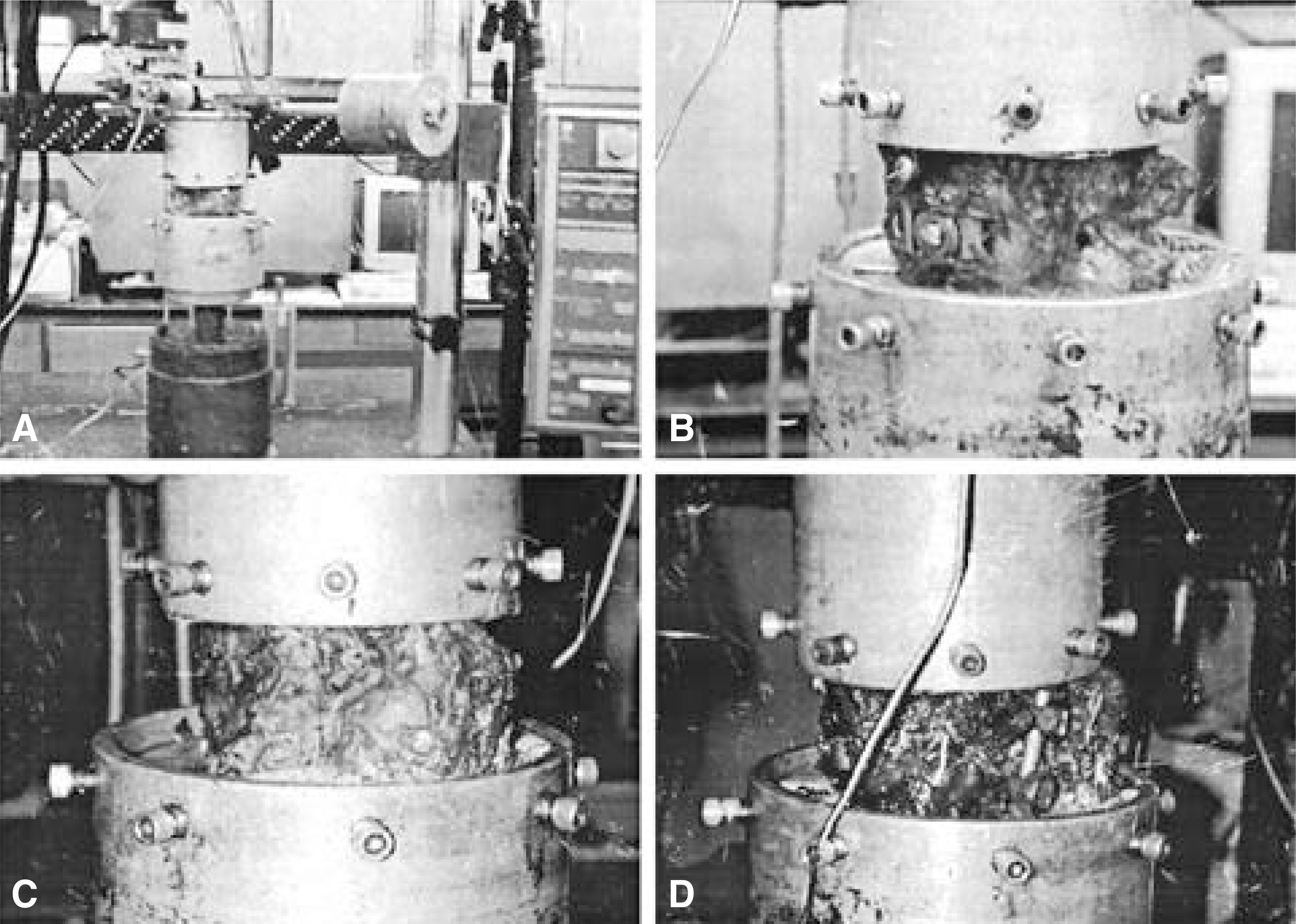
Flexibility was tested through the unconstrained eccentric compression-bending of isolated L4-L5 motion segments. A total of sixteen fresh frozen human cadaveric lumbosacral spine specimens(range of ages : 42+/-13 years 12 males and 4 females) were tested in this investigation. In each case bending load was applied in flexion(0 degree direction), then in 30 degree increments around the transverse plane until flexion was repeated at the 360 degree loading direction. Specimens underwent anterior interbody instrumentation with three different types of cage at L4-5 in three groups, respectively. After testing the interbody fusion constructs, the L4-L5 segments were first stabilized posteriorly using transfacet screws and then retested using transpedicular screw instrumentation.
RESULTS
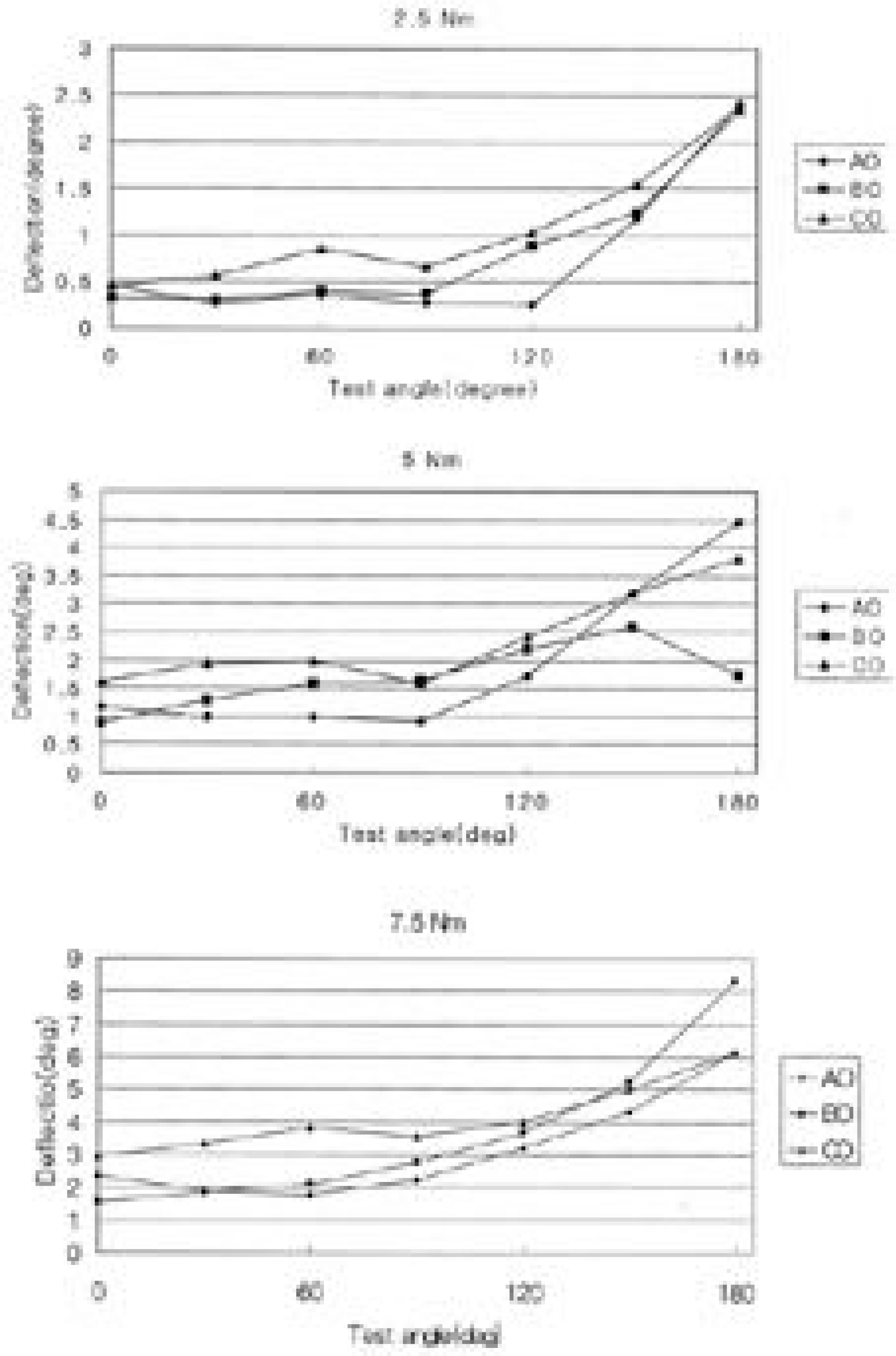
In the intact model, the increase in deflection angle was twice compared with that of the previous point starting from 120 degree up to 150 degree. The pure extensional motion showed the largest deflection angles which are 3.5 times higher than those in pure flexion in average. All three types of cages showed the similar results that were obtained from the intact model.
MeSH Terms
Figure
Reference
-
1). Boden SD, Sumner DR. Biologic factors affecting spinal fusion and bone regeneration. Spine. 20:102S–112S. 1995.
Article2). Brantigan JW, Steffee AD. A carbon fiber implant to aid interbody lumbar fusion. two-year clinical results in the first 2 patients. Spine. 18:2106–7. 1993.3). Bratigan JW, Cunningham BW, Warden K, McAfee PC, Steffee AD. Compression strength of donor bone for posterior lumbar interbody fusion. Spine. 18:1213–1221. 1993.
Article4). Brodke DS, Dick JC, Kunz DN, KcCabe R, Zdeblick TA. Posterior lumbar interbody fusion. Spine. 22:26–31. 1997.
Article5). Cloward R. The treatment of ruptured intervertebral disc by vertebral body fusion. Indication, operation technique after care. J Neurosurg. 10:154–168. 1953.6). Cotterill PC, Kostuik JP, D'Angelo G, Fernie GR, Maki BE. An anatomical comparison of the human and bovine thoracolumbar spine. J Orthop Res. 4:298–303. 1986.
Article7). Deguchi M. Biomechanical evaluation of translaminar facet joint fixation. Acomparative study of poly-L-lactide pins, screws, and pedicle fixation. Spine. 23:1307–1313. 1998.8). Dimar JR, Wang M, Beck DJ, Glassman SD, Voor MJ. Posterior Lumbar Intebody Cages Do Not Agument Segmental Biomechanical Stability. Spine(in-Press).9). Enker P, Steffee AD. Interbody fusion and instrumentation. Clin Orthop. 300:90–101. 1994.
Article10). Evans JH. Biomechanics of lumbar fusion. Clin Orthop Vol. 193:38–46. 1985.
Article11). Fraser RD. Interbody, posterior, and combine lumbar fusion. Spine. 20:167–177. 1995.12). Glazer PA, Colliou O, Lotz JC, Bradford DS. Biomechanical analysis of lumboscral fixation. Spine. 21:1211–1222. 1996.13). Hoshijima K, Nightingale RW, Yu JR, Richardson WJ, Harper KD, Yamamoto H, Myers BS. Strength and stability of posterior lumbar interbody fusion. Comparison of titanium fiber mesh implant and tricortical bone graft. Spine. 22:1181–8. 1997.14). Jost B, Cripton PA, Lund T. Compressive strength of interbody cages in the lumbar spine. The effect of cage shape, posterior instrumentation, and bone density. Eur spine J. 7:132–141. 1998.
Article15). Kozak JA, Heilman AE, O'Brien JP. Anterior lumbar fusion options, Technique and graft materials. Clin Orthop. 300:45–51. 1994.
Article16). Lin PM, Cautilli RA, Joyce MF. Posterior lumbar interbody fusion. Clin Orthop. 180:154–168. 1983.
Article17). Loguidice VA, Johnson RG, Guyer RD. Anterior lumbar interbody fusion. Spine. 13:166–9. 1988.
Article18). Lorentz M, Patwardhan A, Vanderby R Jr. Load-bearing characteristics of lumbar facet in normal and surgically altered spinal segments. Spine. 8:122–130. 1983.19). Lund T, Oxland TR, Jost B, et al. Interbody cage stabilization in the lumbar spine: Evaluation of cage design, posterior instrumentation and bone density. J Bone Joint Surg. 90B:351–9. 1998.20). Lund T, Oxland TR, Jost B, Cripton P, Glassmann S, Etter C, Nolte LP. Interbody cage stabilization in the lumbar spine-biomechnical evaluation of cage design, posterior instrumentation and bone density. J Bone Joint Surg. 80-B(2):351–359. 1998.21). Nibu K, Panjabi MM, Oxland T. Multi-directional stabilizing potential of BAKinterbody fusion system for anterior surgery. J Spinal Disord. 10:357–362. 1997.22). Oxland TR, Zoltan F, Thomas N. A comparative bio -mechanical investigation of anterior lumbar interbody cages-central and bilateral approaches. J Bone Joint Surg(Am). 82-A:383–393. 2000.23). Panjabi MM. Biomechanical evaluation of spinal fixation devices. I. A conceptual framework. Spine. 13:1129–1134. 1998.24). Pfeiffer M, Griss P, Haake M. Standardized evaluation of long term results after anterior lumbar interbody fusion. Eur spine J. 5:299–307. 1996.25). Rapoff AJ, Ghanayem AJ, Zdeblick TA. Biomechanical comparison of posterior lumbar interbody cages. Spine. 22:2375–2379. 1997.26). Soini J. Lumbar disc space heights after external fixation and anterior interbody fusion: A prospective 2-year followup of clinical and radiographic results. J Spinal Disord. 7:487–94. 1994.27). Tencer AF, Hampton D, Eddy S. Biomechanical properties of threadeed inserts for lumbar interbody fusion. Spine. 20:2408–2414. 1995.28). Yang KH, King AI. Mechanism of facet load transmission as a hypothesis for low back pain. Spine. 9:557–565. 1984.
- Full Text Links
- Actions
-
Cited
- CITED
-
- Close
- Share
- Similar articles
-
- Biomechanical Comparison of Multilevel Stand-Alone Lumbar Lateral Interbody Fusion With Posterior Pedicle Screws: An In Vitro Study
- The Change of Segmental Sagittal angle in Low - grade spondylolisthesis after Pedicular Screw Fixation with or without PLIF - PLIF + PLF versus PLF groups -
- Soft GRAF Fixation and Posterior Lumbar Interbody Fusion in Multiple Degenerative Lumbar Diseases
- Comparative Finite Element Analysis of Lumbar Cortical Screws and Pedicle Screws in Transforaminal and Posterior Lumbar Interbody Fusion
- The Effect of Anterior Column Augmentation in Thoraco-lumbar Burst Fractures Treated with Pedicle Screw Instrumentation


