J Korean Soc Spine Surg.
2011 Sep;18(3):169-173.
Delayed Paraplegia after Successful Percutaneous Vertebroplasty in a Patient with Osteoporotic Compression Fracture: A Case Report
- Affiliations
-
- 1Department of Orthopaedic Surgery, Hallym University Sacred Heart Hospital, Medical College of Hallym University, Anyang, Korea.
- 2Department of Orthopaedic Surgery, Ajou University School of Medicine, Suwon, Korea. namsuchung@gmail.com
Abstract
- STUDY DESIGN: A case report.
OBJECTIVES
We report a case of a female patient initially diagnosed as osteoporotic vertebral fracture without any noticeable injuries to posterior ligament complex, who later developed with incomplete paraplegia resulting from an unrecognized trauma after vertebroplasty. SUMMARY OF LITERATURE REVIEW: Vertebroplasty remains a safe and effective procedure for osteoporotic vertebral fracture. However, there have been many reports regarding neural injury associated with cement leakage.
MATERIALS AND METHODS
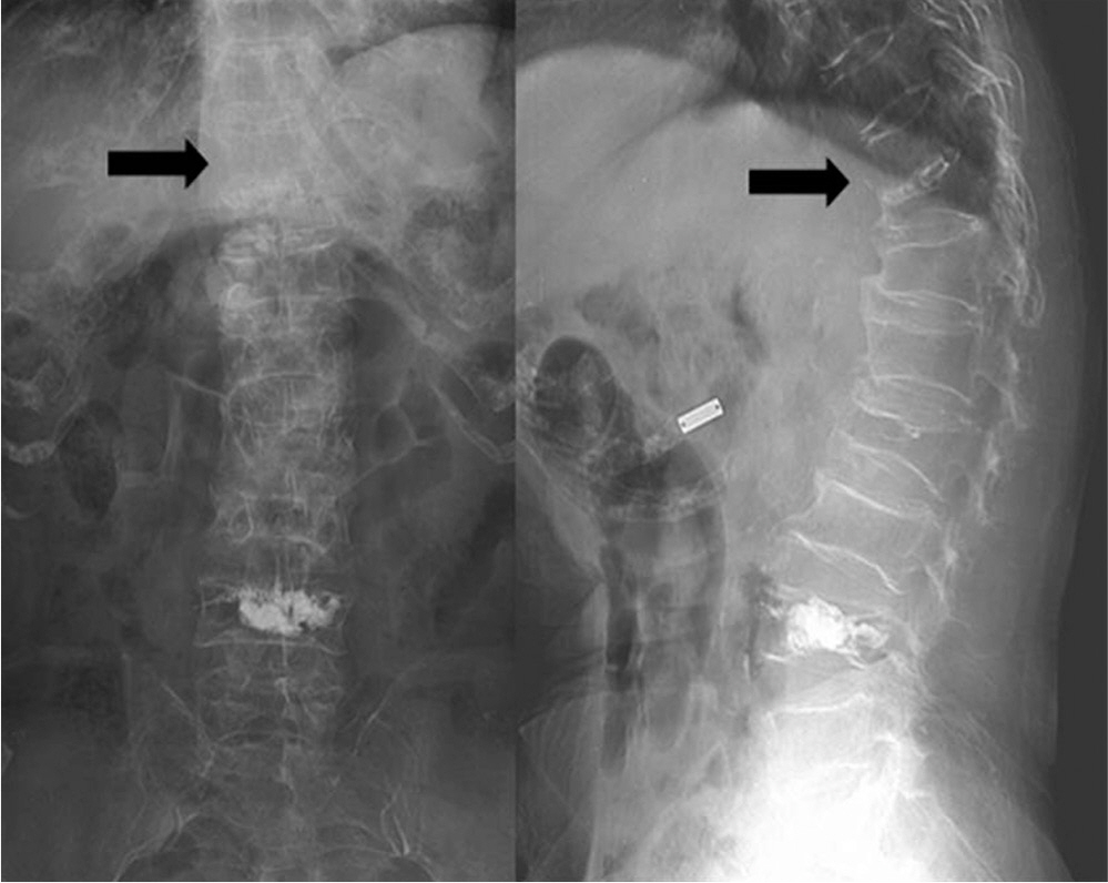
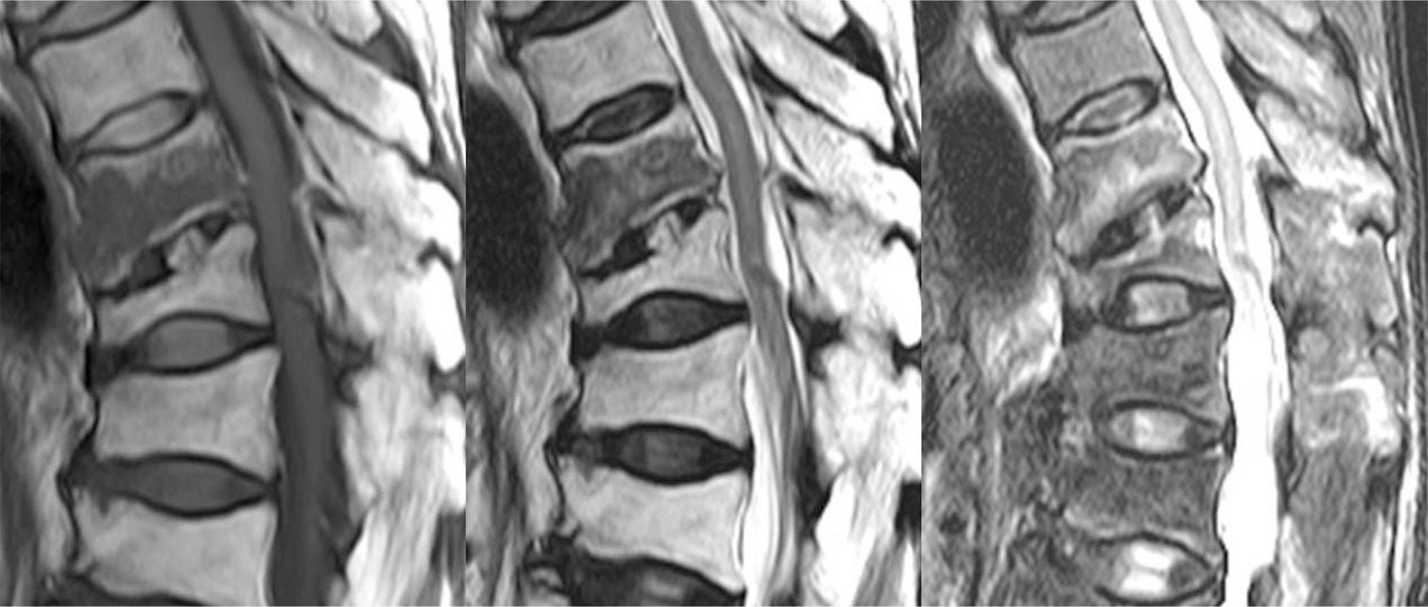
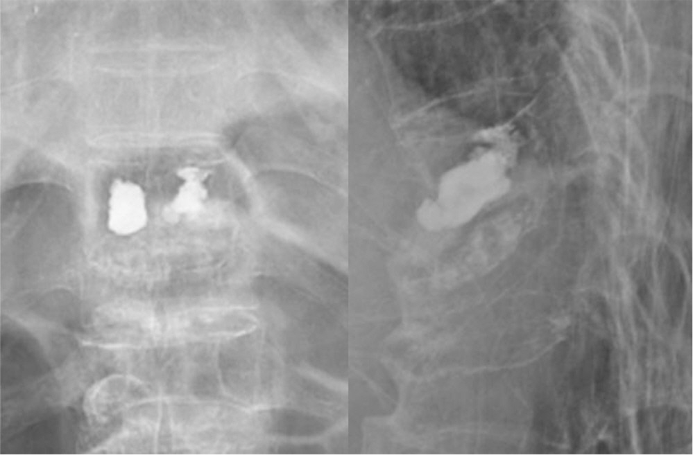
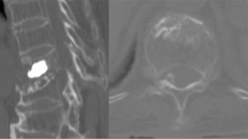
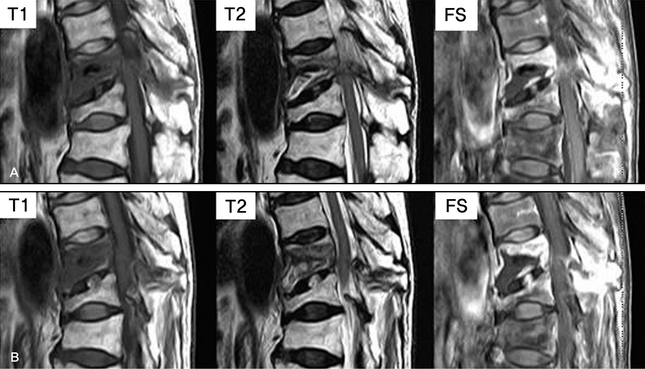
An 81-year old woman with a sudden motor weakness and a sensory loss on her lower extremities after an unrecognized trauma was admitted to our clinic. She had undergone a vertebroplasty twelve days before the admission. At the time of vertebroplasty, Magnetic resonance (MR) imaging showed a compression fracture at T10 vertebra without any posterior ligament complex (PLC) injury. Follow up MR imaging was taken 12 days after vertebroplasty, and it revealed posterior shift of T10 body with a fracture of spinous process, tear of left facet joint capsule, partial tear of interspinous ligament of T10-11 with retrolisthesis, and narrowing of spinal canal at T10-11 by T11 lamina.
RESULTS
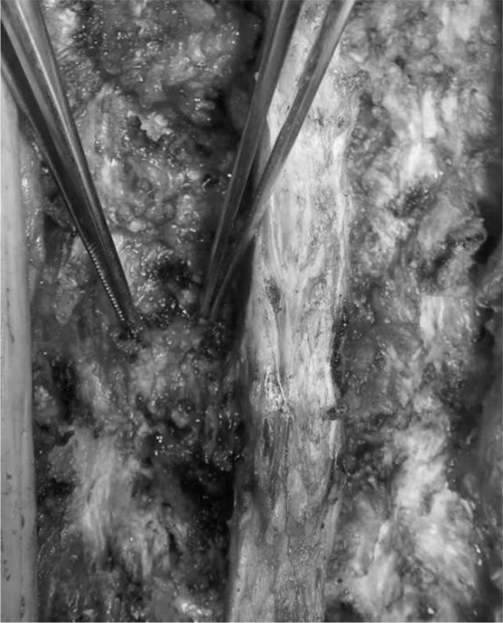
Immediate surgical treatment was performed to decompress the neural structures, and to stabilize the spinal column. However, neurological recovery was unsatisfactory.
CONCLUSIONS
Spinal surgeons should be aware of the possibility of the development of any neurologic deterioration, even if successful vertebroplasty is performed.
Keyword
MeSH Terms
Figure
Reference
-
1. Krueger A, Bliemel C, Zettl R, Ruchholtz S. Management of pulmonary cement embolism after percutaneous vertebroplasty and kyphoplasty: a systematic review of the literature. Eur Spine J. 2009; 18:1257–65.
Article2. Peh WC, Gilula LA. Percutaneous vertebroplasty: indications, contraindications, and technique. Br J Radiol. 2003; 76:69–75.
Article3. Han IH, Chin DK, Kuh SU, et al. Magnetic resonance imaging findings of subsequent fractures after vertebroplasty. Neurosurgery. 2009; 64:740–4.
Article4. Denis F. The three column spine and its significance in the classification of acute thoracolumbar spinal injuries. Spine (Phila Pa 1976). 1983; 8:817–31.
Article5. Oner FC, van Gils AP, Dhert WJ, Verbout AJ. MRI findings of thoracolumbar spine fractures: a categorisation based on MRI examinations of 100 fractures. Skeletal Radiol. 1999; 28:433–43.
Article6. Lee JY, Vaccaro AR, Schweitzer KM Jr, et al. Assessment of injury to the thoracolumbar posterior ligamentous complex in the setting of normal-appearing plain radiography. Spine J. 2007; 7:422–7.
Article7. Lee HM, Kim HS, Kim DJ, Suk KS, Park JO, Kim NH. Reliability of magnetic resonance imaging in detecting posterior ligament complex injury in thoracolumbar spinal fractures. Spine (Phila Pa 1976). 2000; 25:2079–84.
Article8. Petersilge CA, Pathria MN, Emery SE, Masaryk TJ. Thoracolumbar burst fractures: evaluation with MR imaging. Radiology. 1995; 194:49–54.
Article9. Haba H, Taneichi H, Kotani Y, et al. Diagnostic accuracy of magnetic resonance imaging for detecting posterior ligamentous complex injury associated with thoracic and lumbar fractures. J Neurosurg. 2003; 99(1 Suppl):S20–6.
Article
- Full Text Links
- Actions
-
Cited
- CITED
-
- Close
- Share
- Similar articles
-
- Vertebroplasty in the Multiple Osteoporotic Compression Fracture
- Asymptomatic Pulmonary Embolus Following Percutaneous Kyphoplasty: A Case Report
- Large Pulmonary Embolus after Percutaneous Vertebroplasty: A Case Report
- Root Injury after Percutaneous Vertebroplasty in Compression Fracture: Case Report
- The Effect of Early Percutaneous Vertebroplasty in Occult Osteoporotic Vertebral Fracture

