J Korean Soc Radiol.
2011 Sep;65(3):289-295.
Scoliotic Change in Patients Having Undergone a Mastectomy: Analysis Using Multi-Detector Computed Tomography
- Affiliations
-
- 1Department of Radiology, Wonkwang University School of Medicine, Iksan, Korea. khw@wonkwang.ac.kr
Abstract
- PURPOSE
To evaluate the relationship between the degree of scoliotic curvature and postoperative change of the chest wall among patients who underwent a unilateral mastectomy.
MATERIALS AND METHODS
Subjects were comprised of 56 women who underwent chest CT and a whole spine standing anteroposterior view (WSSAP). Cobb's angle and each side of the chest wall volume, including the breast, were measured with the WSSAP and a 3-D reconstructed multi-detector computed tomography (MDCT) image. A correlation analysis was performed between the scoliotic curvature and chest wall volume asymmetry. Directional correspondence between development of scoliosis and undergoing a mastectomy was analyzed. Furthermore, a survey on patient shoulder function was performed using a questionnaire and a correlation was performed between the results of the survey and the scoliotic curvature and chest wall volume asymmetry.
RESULTS
The findings indicate that Cobb's angles were 4.4degrees +/- 2.7 (Mean +/- SD, range from 0.6 to 11.4). Differences in chest wall volume were 474.64 +/- 276.36 cm3 (Mean +/- SD, range from 78 to 1379). No statistical significance was noted between the degree of scoliotic curvature and chest wall volume asymmetry (p > 0.05). A cross-tabulation analysis of the direction between the scoliotic curvature and mastectomy was found to be statistically significant (p < 0.001). Also, there was a significant correlation between shoulder function assessment score and the degree of scoliotic curvature (p = 0.003), while no significant correlation between shoulder function assessment score and chest wall volume asymmetry (p = 0.091) could be found.
CONCLUSION
Scoliotic change had a tendency to be on the opposite side of the mastectomy and had no statistically significant relationship with the volume asymmetry. Thus, 3-D reconstructed MDCT images are helpful in differentiating selective volume differences.
MeSH Terms
Figure
-
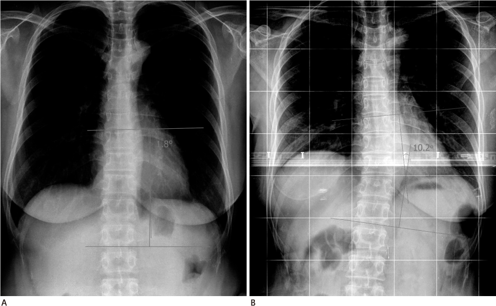
Fig. 1 Measuring Cobb's angle by Cobb method of 44 years old female patient. A. Preoperative chest plane radiograph. B. Whole spine standing anteroposterior view after 4 years.

Fig. 2 Measured volume of the chest wall by 3D reconstructed volume rendering image of chest MDCT. A. Whole chest wall volume is divided into 4 segments by right anterior (RtA), left anterior (LtA), right posterior (RtP), left posterior (LtP). B. The muscle volume of chest wall is measured with 20-80 HU threshold (mRtA, mLtA, mRtP, mLtP). Note.-MDCT = multi-detector computed tomography
Reference
-
1. Jemal A, Siegel R, Ward E, Hao Y, Xu J, Murray T, et al. Cancer Statistics, 2008. CA Cancer J Clin. 2008; 58:71–96.2. Ministry for Health, Welfare and Family Affairs. Annual Report of cancer incidence (2007), cancer prevalence (2007) and survival (1993-2007) in Korea. Seoul: Ministry for Health, Welfare and Family Affairs;2009.3. Won YJ, Sung J, Jung KW, Kong HJ, Park S, Shin HR, et al. Nationwide cancer incidence in Korea, 2003-2005. Cancer Res Treat. 2009; 41:122–131.4. Kuehn T, Klauss W, Darsow M, Regele S, Flock F, Maiterth C, et al. Long-term morbidity following axillary dissection in breast cancer patient--clinical assessment, significance for life quality and the impact of demographic, oncologic and therapeutic factors. Breast Cancer Res Treat. 2000; 64:275–286.5. Bower JE, Ganz PA, Desmond KA, Bernaards C, Rowland JH, Meyerowitz BE, et al. Fatigue in long-term breast carcinoma survivors: a longitudinal investigation. Cancer. 2006; 106:751–758.6. Bendz I, Fagevik Olsén M. Evaluation of immediate versus delayed shoulder exercise after breast cancer surgery including lymph node dissection--a randomised controlled trial. Breast. 2002; 11:241–248.7. Wingate L. Efficacy of physical therapy for patients who have undergone mastectomies. A prospective study. Phys Ther. 1985; 65:896–900.8. Aebi M. The adult scoliosis. Eur Spine J. 2005; 14:925–948.9. Lee SW, Lee TJ, Lee SW. Does unilateral mastectomy cause scoliosis? J Korean Soc Plast Reconstr Surg. 2008; 35:279–282.10. Shamley D, Srinaganathan R, Oskrochi R, Lascurain-Aguirrebeña I, Sugden E. Three-dimensional scapulothoracic motion following treatment for breast cancer. Breast Cancer Res Treat. 2009; 118:315–322.11. Cheville AL, Tchou J. Barriers to rehabilitation following surgery for primary breast cancer. J Surg Oncol. 2007; 95:409–418.12. Hagay C, Cherel PJ, de Maulmont CE, Plantet MM, Gilles R, Floiras JL, et al. Contrast-enhanced CT: value for diagnosing local breast cancer recurrence after conservative treatment. Radiology. 1996; 200:631–638.13. Lindfors KK, Meyer JE, Busse PM, Kopans DB, Munzenrider JE, Sawicka JM. CT evaluation of local and regional breast cancer recurrence. AJR Am J Roentgenol. 1985; 145:833–837.
- Full Text Links
-
- Actions
-
Cited
- CITED
-
- Close
- Share
-
- Similar articles
-
- Three cases of right coronary anomaly confirmed by multi-detector computed tomography
- Overlooked or unrecognized pitfalls in noninvasive multi-detector computed tomography coronary angiography
- Comparative evaluation of computed tomography for dental implants on the mandibular edentulous area
- Spectrum of Multi-Detector Computed Tomography Findings that Alter Pulmonary Artery Diameters in Adults
- Multi-Detector Row CT of the Central Airway Disease
