Duplication of the Left Vertebral Artery Origin: A Case Report
- Affiliations
-
- 1Department of Radiology, College of Medicine, Hanyang University, Hanyang University Guri Hospital, Guri, Korea. dwpark@hanyang.ac.kr
Abstract
- Duplication of vertebral arteries is a very rare but clinically important condition. A duplicated vertebral artery origin can influence hemodynamics, pathogenesis of vascular lesions and treatment options. In cases of vertebral artery duplication, the vertebral arteries generally enter the transverse foramen higher up than normal. Awareness of these vertebral artery variants before procedures, such as neurointervention or surgery, may be beneficial. Here, we describe a case of a 51-year-old female patient with left vertebral artery duplication which was detected incidentally.
MeSH Terms
Figure
-
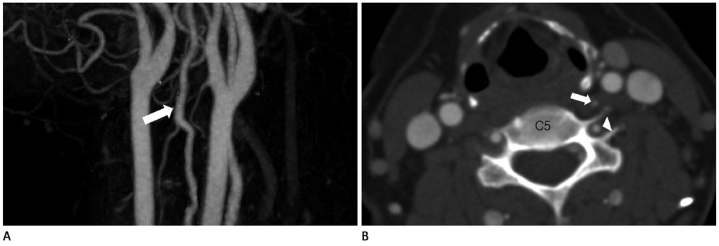
Fig. 1 Duplication of the left vertebral artery origin in a 51-year-old female patient (magnetic resonance angiogram not shown). A. Computed tomography angiography (CTA) maximum intensity projection image shows that the duplicated limbs of the left vertebral artery unite to form the distal part of the left vertebral artery (arrow). B. CTA axial source image shows a hypoplastic limb (arrow) arising from the aortic arch and a normal limb (arrowhead) arising from the left subclavian artery at the C5 level.

Fig. 2 Duplication of the left vertebral artery origin in a 51-year-old female patient. A. Left subclavian angiographic image shows the normal limb of the duplicated left vertebral artery (arrow) arising from the left subclavian artery. B, C. Selective angiography of the anomalous hypoplastic limb (arrowhead) shows it is the third branch of the aortic arch, and opacifies the left vertebral artery as a result of the retrograde flow into the normal limb (arrow).
Reference
-
1. Matula C, Trattnig S, Tschabitscher M, Day JD, Koos WT. The course of the prevertebral segment of the vertebral artery: anatomy and clinical significance. Surg Neurol. 1997. 48:125–131.2. Satti SR, Cerniglia CA, Koenigsberg RA. Cervical vertebral artery variations: an anatomic study. AJNR Am J Neuroradiol. 2007. 28:976–980.3. Koenigsberg RA, Pereira L, Nair B, McCormick D, Schwartzman R. Unusual vertebral artery origins: examples and related pathology. Catheter Cardiovasc Interv. 2003. 59:244–250.4. Kendi AT, Brace JR. Vertebral artery duplication and aneurysms: 64-slice multidetector CT findings. Br J Radiol. 2009. 82:e216–e218.5. Goddard AJ, Annesley-Williams D, Guthrie JA, Weston M. Duplication of the vertebral artery: report of two cases and review of the literature. Neuroradiology. 2001. 43:477–480.6. Panicker HK, Tarnekar A, Dhawane V, Ghosh SK. Anomalous origin of left vertebral artery - embryological basis and applied aspects - A case report. J Anat Soc India. 2002. 51:234–235.7. Bergman RA, Afifi AF, Miyauchi R. Illustrated Encyclopedia of Human Anatomic Variation. Available at: http://www.anatomyatlases.org/AnatomicVariants/Cardiovsacular/Images0001/0095.shtml.8. Bruneau M, Cornelius JF, Marneffe V, Triffaux M, George B. Anatomical variations of the V2 segment of the vertebral artery. Neurosurgery. 2006. 59:1 Suppl 1. ONS20–ONS24. discussion ONS20-ONS24.9. Goray VB, Joshi AR, Garg A, Merchant S, Yadav B, Maheshwari P. Aortic arch variation: a unique case with anomalous origin of both vertebral arteries as additional branches of the aortic arch distal to left subclavian artery. AJNR Am J Neuroradiol. 2005. 26:93–95.10. Komiyama M, Morikawa T, Nakajima H, Nishikawa M, Yasui T. High incidence of arterial dissection associated with left vertebral artery of aortic origin. Neurol Med Chir (Tokyo). 2001. 41:8–11. discussion 11-12.
- Full Text Links
-
- Actions
-
Cited
- CITED
-
- Close
- Share
-
- Similar articles
-
- Duplicated Origin of the Left Vertebral Artery: A Case Report and Embryological Review
- Duplicated Vertebral Artery : Literature Review and Clinical Significance
- Pseudocoarctation of the Aorta Associated with the Anomalous Origin of the Left Vertebral Artery: a Case Report
- Aberrant Right Vertebral Artery Originating from the Aortic Arch Distal to the Left Subclavian Artery: A Case Report
- Two Cases of Aberrant Vertebral Artery Originating from Aortic Arch Distal to Left Subclavian Artery
