Subcutaneous Cavernous and Capillary Hemangiomas of the Breast: Radiologic-Pathological Correlation
- Affiliations
-
- 1Department of Radiology, Soonchunhyang University Cheonan Hospital, Cheonan, Korea. taloo@hanmail.net
- 2Department of Pathology, Soonchunhyang University Cheonan Hospital, Cheonan, Korea.
Abstract
- Vascular tumors of the breast are uncommon and include hemangioma, angiolipoma, and angiosarcoma. Among breast hemangiomas, capillary hemangiomas are relatively rare in contrast to cavernous hemangiomas. We report two rare cases of cavernous and capillary hemangiomas of the breast by mammography, sonography, and positron emission tomography/computed tomography.
MeSH Terms
Figure
-
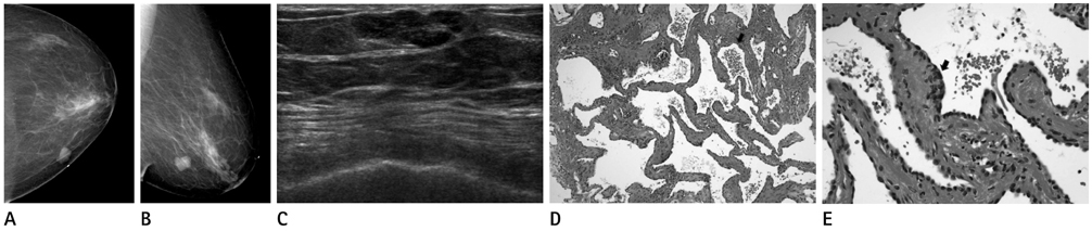
Fig. 1 An 80-year-old woman with cavernous hemangioma in left breast. A, B. Craniocaudal and mediolateral oblique mammograms show a well-circumscribed lobular isodense nodule at palpable site. C. Transverse sonography shows a well-circumscribed lobular hypoechoic nodule in subcutaneous fat layer. D. Microscopic examination reveals dilated vessels (arrow) congested with red blood cells (H&E stain, × 40). E. Numerous large vascular channels that share a common wall and lined with a single layer of endothelial cells (arrow) without any atypical cells, suggesting cavernous hemangioma (H&E stain, × 100).
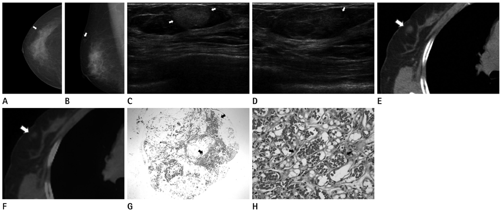
Fig. 2 A 72-year-old woman with two capillary hemangiomas in right breast. A, B. The patient presented with a palpable mass in the right breast upper inner quadrant. Craniocaudal and mediolateral oblique mammograms show no abnormality at the palpable site. A partly obscured oval isodense nodule (arrows) was incidentally detected in the right breast upper outer quadrant. C. Transverse sonography of right upper outer breast shows a circumscribed oval hyperechoic nodule (arrows) in subcutaneous fat layer. D. Transverse sonography of right inner breast shows a palpable partly indistinct oval mixed isoechoic nodule (arrow) in subcutaneous fat layer. E. Nonfused CT image shows a partly ill-defined oval isodense nodule (arrow) in right upper outer breast. Another palpable nodule in right upper inner breast was invisible. F. Fused PET/CT image demonstrates no definite increased FDG uptake at nodule (arrow) in right upper outer breast. G. Excisional biopsy was also performed for two nodules. Microscopic examination of palpable nodule in right upper inner breast reveals infiltrative tumor (arrows) into adjacent adipose tissue (H&E stain, × 12.5). Another nonpalpable nodule had a similar microscopic feature (not shown in figure). H. It is composed of congested very small blood vessels that resemble capillaries (arrows), lined with a single layer of endothelial cells (H&E stain, × 400). Note.-FDG = fluoro-2-deoxy-D-glucose, PET/CT = positron emission tomography/computed tomography
Reference
-
1. Mesurolle B, Sygal V, Lalonde L, Lisbona A, Dufresne MP, Gagnon JH, et al. Sonographic and mammographic appearances of breast hemangioma. AJR Am J Roentgenol. 2008; 191:W17–W22.2. Siewert B, Jacobs T, Baum JK. Sonographic evaluation of subcutaneous hemangioma of the breast. AJR Am J Roentgenol. 2002; 178:1025–1027.3. Choi KH, Chang YW, Yang SB. Subcutaneous cavernous hemangioma of the breast: a case report. J Korean Radiol Soc. 2008; 59:209–211.4. Lee HB, Kim JY, Kim SH, Jeong MJ, Kim JH, Kim SH, et al. Cavernous hemangioma of the breast as a palpable mass: a case report. J Korean Soc Radiol. 2009; 60:207–209.5. Vieira SC, Silva JS, Madeira EB, França JCQ, Martins Filho SN. Breast hemangioma mimicking metastasis at PET-CT. Radiol Bras. 2011; 44:401–402.6. Hayasaka K, Tanaka Y, Saitoh T, Takahashi M. Gadolinium-enhanced dynamic MRI of breast hemangioma. Comput Med Imaging Graph. 2003; 27:493–495.7. Rosen PP, Jozefczyk MA, Boram LH. Vascular tumors of the breast. IV. The venous hemangioma. Am J Surg Pathol. 1985; 9:659–665.8. Schäfer FK, Biernath-Wuepping J, Eckmann-Scholz C, Order BM, Mathiak M, Hilpert F, et al. Rare Benign Entities of the Breast - Myoid Hamartoma and Capillary Hemangioma. Geburtsh Frauenheilk. 2012; 72:412–418.9. Rosen PP. Rosen's Breast Pathology. 3rd ed. Philadelphia: Lippincott Williams & Wilkins;2009. p. 789–797.10. Pui MH, Movson IJ. Fatty tissue breast lesions. Clin Imaging. 2003; 27:150–155.
- Full Text Links
-
- Actions
-
Cited
- CITED
-
- Close
- Share
-
- Similar articles
-
- Subcutaneous Cavernous Hemangioma of the Breast: A Case Report
- Immediate Breast Reconstruction after Resection of Cavernous Hemangioma
- Cavernous Hemangioma of the Breast as a Palpable Mass: A Case Report
- A Case of Cavernous Hemangioma Originated from the Middle Turbinate
- A Case of Subcutaneous Cavernous Hemangioma Presenting as a Nasolabial Fold Mass
