Undifferentiated Embryonal Sarcoma of the Liver in an Adult: The Verification of the High Growth Rate in the Tumor
- Affiliations
-
- 1Department of Radiology, Daegu Fatima Hospital, Daegu, Korea. praia-zorlborn@hanmail.net
Abstract
- We report the radiologic findings of a rare case of undifferentiated embryonal sarcoma of liver (UES) in a 64-year-old female. To our knowledge, this is the unique case of adult UES that provide verification of the high growth rate of the tumor.
Figure
-
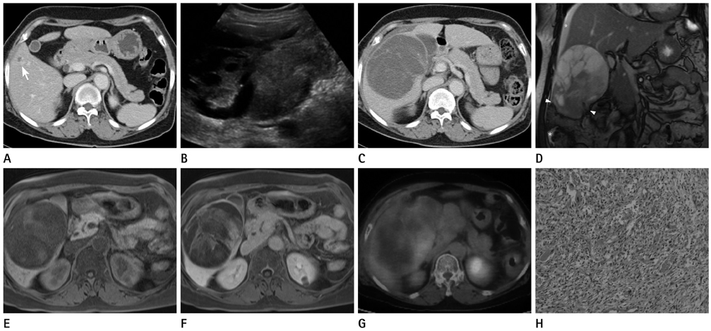
Fig. 1 A 64-year female with undifferentiated embryonal sarcoma of the liver. A. 7 months ago, abdominal CT showed a 0.4 cm hypodense lesion (arrow) in the segment 5 of the liver. B. Transverse US images show the huge and predominantly solid mass in the liver with peripheral cystic components. C. Contrast enhanced CT shows the 11.0 cm-sized hypoattenuating mass in the right lobe of liver with multiple thick septa. D. Coronal T2-weighted MR images show the large heterogeneous mass in the right lobe of the liver. Upper portion of the tumor is hyperintense with septation and lower portion is hypointense. In the lowest part of the mass, rupture (arrowheads) with asscociated intraperitoneal fluid (asterisk) is shown. E, F. Unenhanced T1-weighted and gadolinium contrast enhanced T1-weighted MR images show progressive enhancement of the periphery and septa. G. PET-CT shows diffuse mild FDG uptake in the peripheral portion of the mass. H. Photomicrograph (original magnification, × 200; H&E stain) shows undifferentiated spindle-shaped and large stellate cells in abundant myxoid stroma. Note.-FDG = fluorodeoxyglucose, PET = positron emission tomography, US = ultrasonography
Reference
-
1. Stocker JT, Ishak KG. Undifferentiated (embryonal) sarcoma of the liver: report of 31 cases. Cancer. 1978; 42:336–348.2. Li XW, Gong SJ, Song WH, Zhu JJ, Pan CH, Wu MC, et al. Undifferentiated liver embryonal sarcoma in adults: a report of four cases and literature review. World J Gastroenterol. 2010; 16:4725–4732.3. Buetow PC, Buck JL, Pantongrag-Brown L, Marshall WH, Ros PR, Levine MS, et al. Undifferentiated (embryonal) sarcoma of the liver: pathologic basis of imaging findings in 28 cases. Radiology. 1997; 203:779–783.4. Ros PR, Olmsted WW, Dachman AH, Goodman ZD, Ishak KG, Hartman DS. Undifferentiated (embryonal) sarcoma of the liver: radiologic-pathologic correlation. Radiology. 1986; 161:141–145.5. Joshi SW, Merchant NH, Jambhekar NA. Primary multilocular cystic undifferentiated (embryonal) sarcoma of the liver in childhood resembling hydatid cyst of the liver. Br J Radiol. 1997; 70:314–316.6. Martí-Bonmatí L, Ferrer D, Menor F, Galant J. Hepatic mesenchymal sarcoma: MRI findings. Abdom Imaging. 1993; 18:176–179.7. Psatha EA, Semelka RC, Fordham L, Firat Z, Woosley JT. Undifferentiated (embryonal) sarcoma of the liver (USL): MRI findings including dynamic gadolinium enhancement. Magn Reson Imaging. 2004; 22:897–900.8. Vermess M, Collier NA, Mutum SS, Crofton ME. Misleading appearance of a rare malignant liver tumour on computed tomography. Br J Radiol. 1984; 57:262–265.9. Baron PW, Majlessipour F, Bedros AA, Zuppan CW, Ben-Youssef R, Yanni G, et al. Undifferentiated embryonal sarcoma of the liver successfully treated with chemotherapy and liver resection. J Gastrointest Surg. 2007; 11:73–75.
- Full Text Links
-
- Actions
-
Cited
- CITED
-
- Close
- Share
-
- Similar articles
-
- Embryonal Sarcoma of the Liver in an Adult
- Erratum: Undifferentiated Embryonal Sarcoma of the Liver in an Adult: The Verification of the High Growth Rate in the Tumor
- Undifferentiated embryonal sarcoma of the liver in an adult patient
- Undifferentiated Sarcoma of the Liver in Adult: A Case Report and Review of the Literature
- Undifferentiated Embryonal Sarcoma of Liver in Child
