Perirenal Lymphangiomatosis
- Affiliations
-
- 1Department of Urology, Pusan National University Hospital, Busan, Korea. hongkooha@naver.com
- 2Department of Radiology, Pusan National University Hospital, Busan, Korea.
- 3Department of Pathology, Pusan National University Hospital, Busan, Korea.
- 4Department of Urology, Dong-A University Medical Center, Busan, Korea.
- KMID: 2320803
- DOI: http://doi.org/10.5534/wjmh.2014.32.2.116
Abstract
- Lymphangioma is a rare, benign mesenchymal neoplasm, which is characterized by numerous intercommunicating cystic spaces containing lymphatic fluid. It is considered a congenital disease resulting from the obstruction of regional lymph drainage during the developmental period. Lymphangioma may be focal/unilateral or diffuse/bilateral, and in the latter case, it is referred to as lymphangiomatosis. Here, we report a case of a 38-year-old man with perirenal lymphangiomatosis. The patient's chief complaint was left flank pain, and left pleural effusion was found on radiological examination. After radical nephrectomy, the pathological examinations revealed that the kidney was enclosed by a multicystic mass with intrarenal cystic dilatations. We report the case and discuss the management of perirenal lymphangiomatosis with a literature review.
Keyword
MeSH Terms
Figure
-
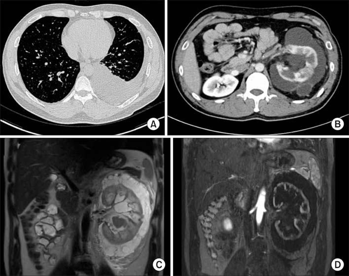
Fig. 1 Radiologic images of perirenal lymphangiomatosis. (A) Chest computed tomography (CT) reveals pleural effusion of the left pleural cavity. (B) An enlarged left kidney has decreased radio-contrast enhancement with septated perirenal cystic fluid collections on abdominal CT. (C) High signal intensity is found around the left kidney with enlarged pelvo-calyceal system on T1-weighted magnetic resonance (MR). (D) Cortical thinning and irregularity of the left kidney is demonstrated in contrast enhanced T2-weighted MR.
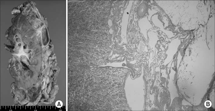
Fig. 2 Gross and microscopic findings. (A) Multilocular cystic mass is enclosing the kidney. The mass consists of multiple cysts having thin fibrous walls and contains clear serous fluid. (B) Low power (×40) microscopic view shows multiloculated cystic spaces with various caliber of muscular and fibrous walls and without definite epithelial lining on H&E staining.
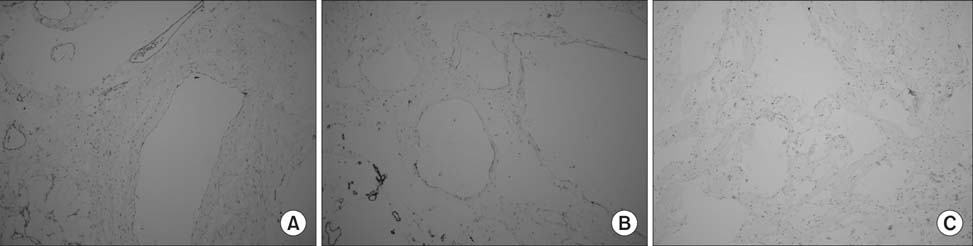
Fig. 3 Flattened endothelial lining is visualized by immunohistochemical staining for CD31 (A), CD34 (B), and HMB-45 (C) (A~C: ×100).
Reference
-
1. Westphalen A, Yeh B, Qayyum A, Hari A, Coakley FV. Differential diagnosis of perinephric masses on CT and MRI. AJR Am J Roentgenol. 2004; 183:1697–1702.
Article2. Lindsey JR. Lymphangiectasia simulating polycystic disease. J Urol. 1970; 104:658–662.
Article3. Honma I, Takagi Y, Shigyo M, Sunaoshi K, Wakabayashi J, Harada O, et al. Lymphangioma of the kidney. Int J Urol. 2002; 9:178–182.
Article4. Leder RA. Genitourinary case of the day. Renal lymphangiomatosis. AJR Am J Roentgenol. 1995; 165:197–198.
Article5. Murray KK, McLellan GL. Renal peripelvic lymphangiectasia: appearance at CT. Radiology. 1991; 180:455–456.
Article6. Varela JR, Bargiela A, Requejo I, Fernandez R, Darriba M, Pombo F. Bilateral renal lymphangiomatosis: US and CT findings. Eur Radiol. 1998; 8:230–231.
Article7. Laurent F, Joullie M, Biset JM, Simon JM, Drouillard J. Cystic lymphangioma of the kidney: a rare cause of multiloculated renal masses. Eur J Radiol. 1991; 12:67–68.
Article8. Kutcher R, Mahadevia P, Nussbaum MK, Rosenblatt R, Freed S. Renal peripelvic multicystic lymphangiectasia. Urology. 1987; 30:177–179.
Article9. Cutillo DP, Swayne LC, Cucco J, Dougan H. CT and MR imaging in cystic abdominal lymphangiomatosis. J Comput Assist Tomogr. 1989; 13:534–536.
Article
- Full Text Links
-
- Actions
-
Cited
- CITED
-
- Close
- Share
-
- Similar articles
-
- Perirenal Lymphangioma Combined With Multiple Splenic and Hepatic Cysts
- Metachronous Bilateral Renal Lymphangiomatosis Mimicking as a Simple Renal Cyst
- Colonic Lymphangiomatosis with Normal Colonoscopic Finding in an Adult
- Lymphangiomatosis of Bone and Soft Tissue: A Case Report
- A Case Report with Lymphangiomatosis of the Colon
