Tuberc Respir Dis.
2015 Jul;78(3):297-301. 10.4046/trd.2015.78.3.297.
Malignant Mesothelioma Diagnosed by Bronchoscopic Biopsy
- Affiliations
-
- 1Department of Internal Medicine, Chungnam National University School of Medicine, Daejeon, Korea. vov-x@daum.net
- KMID: 2320661
- DOI: http://doi.org/10.4046/trd.2015.78.3.297
Abstract
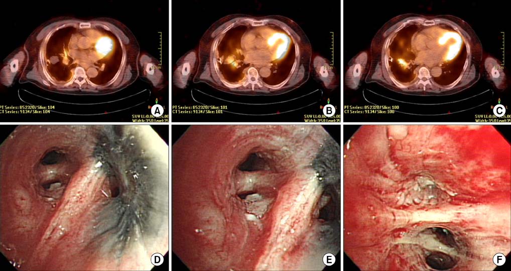
- Malignant mesothelioma is a rare malignant neoplasm that arises from mesothelial surfaces of the pleural cavity, peritoneal cavity, tunica vaginalis, or pericardium. Typically, pleural fluid cytology or closed pleural biopsy, surgical intervention (video thoracoscopic biopsy or open thoracotomy) is conducted to obtain pleural tissue specimens. However, endobronchial lesions are rarely seen and cases diagnosed from bronchoscopic biopsy are also rarely reported. We reported the case of a 77-year-old male who was diagnosed as malignant mesothelioma on bronchoscopic biopsy from obstructing masses of the endobronchial lesion.
Keyword
Figure
Reference
-
1. Price B, Ware A. Time trend of mesothelioma incidence in the United States and projection of future cases: an update based on SEER data for 1973 through 2005. Crit Rev Toxicol. 2009; 39:576–588.2. Tsao AS, Wistuba I, Roth JA, Kindler HL. Malignant pleural mesothelioma. J Clin Oncol. 2009; 27:2081–2090.3. Kim HR, Ahn YS, Jung SH. Epidemiologic characteristics of malignant mesothelioma in Korea. J Korean Med Assoc. 2009; 52:449–455.4. Carteni G, Manegold C, Garcia GM, Siena S, Zielinski CC, Amadori D, et al. Malignant peritoneal mesothelioma. Results from the International Expanded Access Program using pemetrexed alone or in combination with a platinum agent. Lung Cancer. 2009; 64:211–218.5. Mirarabshahii P, Pillai K, Chua TC, Pourgholami MH, Morris DL. Diffuse malignant peritoneal mesothelioma: an update on treatment. Cancer Treat Rev. 2012; 38:605–612.6. Chekol SS, Sun CC. Malignant mesothelioma of the tunica vaginalis testis: diagnostic studies and differential diagnosis. Arch Pathol Lab Med. 2012; 136:113–117.7. Sterman DH, Albelda SM. Advances in the diagnosis, evaluation, and management of malignant pleural mesothelioma. Respirology. 2005; 10:266–283.8. Gadgeel SM, Pass HI. Novel combinations using pemetrexed in malignant mesothelioma. Clin Lung Cancer. 2004; 5:Suppl 2. S61–S66.9. Scherpereel A, Astoul P, Baas P, Berghmans T, Clayson H, de Vuyst P, et al. Guidelines of the European Respiratory Society and the European Society of Thoracic Surgeons for the management of malignant pleural mesothelioma. Eur Respir J. 2010; 35:479–495.10. Kao SC, Yan TD, Lee K, Burn J, Henderson DW, Klebe S, et al. Accuracy of diagnostic biopsy for the histological subtype of malignant pleural mesothelioma. J Thorac Oncol. 2011; 6:602–605.11. Greillier L, Cavailles A, Fraticelli A, Scherpereel A, Barlesi F, Tassi G, et al. Accuracy of pleural biopsy using thoracoscopy for the diagnosis of histologic subtype in patients with malignant pleural mesothelioma. Cancer. 2007; 110:2248–2252.12. Hamamoto J, Notsute D, Tokunaga K, Sasaki J, Kojima K, Saeki S, et al. Diagnostic usefulness of endobronchial ultrasoundguided transbronchial needle aspiration in a case with malignant pleural mesothelioma. Intern Med. 2010; 49:423–426.13. Kang B, Kim MA, Lee BY, Yoon H, Oh DK, Hwang HS, et al. Malignant pleural mesothelioma diagnosed by endobronchial ultrasound-guided transbronchial needle aspiration. Tuberc Respir Dis. 2013; 74:74–78.14. Husain AN, Colby T, Ordonez N, Krausz T, Attanoos R, Beasley MB, et al. Guidelines for pathologic diagnosis of malignant mesothelioma: 2012 update of the consensus statement from the International Mesothelioma Interest Group. Arch Pathol Lab Med. 2013; 137:647–667.15. DellaGiustina D, Falconieri G, Bonifacio-Gori D, Zanconati F, DiBonito L, Pizzolitto S. Bronchial cytology in pleural mesothelioma: a report of 3 positive cases, including 1 diagnosed initially on bronchial brushings. Acta Cytol. 2003; 47:1017–1022.
- Full Text Links
- Actions
-
Cited
- CITED
-
- Close
- Share
- Similar articles
-
- A Case of Malignant Mesothelioma with Pleural Effusion
- Can BAP1 expression loss in mesothelial cells be an indicator of malignancy?
- Two cases of malignant mesothelioma of the peritoneum and pericardium
- Fine needle aspiration cytology of malignant epithelial mesothelioma of the peritoneum
- A Case Report Malignant Mesothelima Diagnosed in the Early Stage by Thoracoscopic Biopsy


