Fistula Formation between Right Upper Bronchus and Bronchus Intermedius Caused by Endobronchial Tuberculosis: A Case Report
- Affiliations
-
- 1Department of Internal Medicine, Chungbuk National University College of Medicine, Cheongju, Korea. drahnjy@chungbuk.ac.kr
- 2Department of Radiology, Chungbuk National University College of Medicine, Cheongju, Korea.
- KMID: 2320658
- DOI: http://doi.org/10.4046/trd.2015.78.3.286
Abstract
- Endobronchial tuberculosis is defined as a tuberculous infection of the tracheobronchial tree and has a prevalence of up to 50% in active pulmonary tuberculosis cases. The most common complication of endobronchial tuberculosis is bronchial stenosis; benign fistula formation by endobronchial tuberculosis is rare, especially inter-bronchial fistula formation. We reported a rare case of a 73-year-old woman with a fistula between the right upper bronchus and bronchus intermedius. A diagnosis of inter-bronchial fistula caused by endobronchial tuberculosis was based on the results of chest computed tomography scans, bronchoscopy, and microbiological and pathological tests. The patient was treated with anti-tuberculous medication, and her symptoms gradually improved.
MeSH Terms
Figure
-
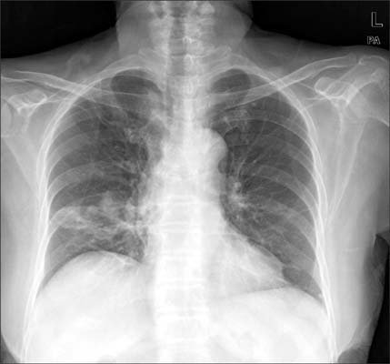
Figure 1 A chest radiograph revealed an ill-defined consolidation in the right lower lung fields and fibrotic change in the right upper lung fields.
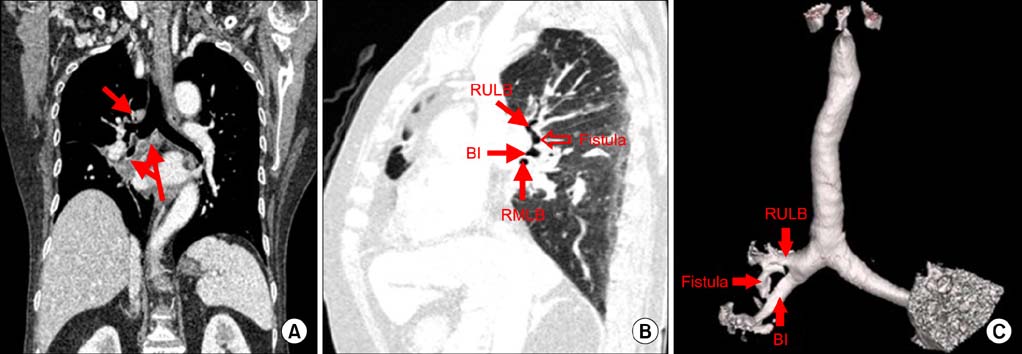
Figure 2 (A) Coronal image of the chest obtained using computed tomography (CT) scan revealed multiple variable-sized mediastinal lymph nodes. (B) Sagittal image of chest obtained using CT scan showed the whole pathway of the broncho-bronchial fistula, which originated in the right upper lobe bronchus and inserted in the bronchus intermedius. (C) The volume rendering image showed the presence of a fistula between the right upper lobe bronchus and the bronchus intermedius. RULB: right upper lobe bronchus; BI: bronchus intermedius; RMLB: right middle lobe bronchus.

Figure 3 (A) A round bronchial wall defect of approximately 3 mm was found at the anterior wall of the right upper lobar bronchus. (B) A round bronchial wall defect of approximately 3 mm was found at the lateral aspect of the distal bronchus intermedius. RULB: right upper lobe bronchus; RLLB: right lower lobe bronchus; RMLB: right middle lobe bronchus.
Reference
-
1. Jung SS, Park HS, Kim JO, Kim SY. Incidence and clinical predictors of endobronchial tuberculosis in patients with pulmonary tuberculosis. Respirology. 2015; 20:488–495.2. Donath J, Khan FA. Tuberculous and posttuberculous bronchopleural fistula. Ten year clinical experience. Chest. 1984; 86:697–703.3. Yilmaz E, Akkoclu A, Sevinc C. CT and MRI appearance of a fistula between the right and left main bronchus caused by tracheobronchial tuberculosis. Br J Radiol. 2001; 74:1056–1058.4. Rikimaru T. Endobronchial tuberculosis. Expert Rev Anti Infect Ther. 2004; 2:245–251.5. Xue Q, Wang N, Xue X, Wang J. Endobronchial tuberculosis: an overview. Eur J Clin Microbiol Infect Dis. 2011; 30:1039–1044.6. Nanaware SK, Gothi D, Joshi JM. Tuberculous broncho-esophageal fistula managed conservatively. Lung India. 2005; 22:65–67.7. Sarkar P, Chandak T, Shah R, Talwar A. Diagnosis and management bronchopleural fistula. Indian J Chest Dis Allied Sci. 2010; 52:97–104.8. Choi YI, Cho JH, Shim JY, Sheen SS, Oh YJ, Park JH, et al. A case of RUL bronchopleural fistula occluded by flexible bronchoscope with endobronchial watanabe spigot (EWS). Tuberc Respir Dis. 2005; 58:404–409.
- Full Text Links
-
- Actions
-
Cited
- CITED
-
- Close
- Share
-
- Similar articles
-
- Reconstruction of Tracheobronchial Stenosis due to Endobronchial Tuberculosis : A case report
- A Case of Displaced Lobar Tracheal Bronchus Associated with Bronchiectasis
- Middle Lobe Syndrome Caused by Occult Foreign Body in the Bronchus Intermedius: A case report
- Right Lower Sleeve Bilobectomy for Lung Cancer with Posteparterial Tracheal Bronchus
- Accessory Cardiac Bronchus with Lung Cancer: A case report
