Tuberc Respir Dis.
2011 Jun;70(6):511-515.
A Rare Case of Fat-Forming Variant of Solitary Fibrous Tumor Presenting as a Pleural Mass
- Affiliations
-
- 1Department of Internal Medicine, Bundang CHA Hospital, CHA University College of Medicine, Seongnam, Korea. imekkim@hanmail.net
- 2Department of Pathology, Bundang CHA Hospital, CHA University College of Medicine, Seongnam, Korea.
- 3Department of Thoracic and Cardiovascular Surgery, Bundang CHA Hospital, CHA University College of Medicine, Seongnam, Korea.
Abstract
- The fat-forming variant of solitary fibrous tumors (SFTs) is a rare soft tissue neoplasm that was previously referred to as a lipomatous hemangiopericytoma (L-HPC). The most common affected site is deep soft tissue. Here, we present the first case, worldwide, of a fat-forming variant of SFT of the pleura. A 74-year-old man presented with left lower chest pain. Chest radiographs showed a mass-like lesion at the left lower lung field and chest computed tomography revealed a 12 cm fat-containing enhancing mass that was well-separated, lobulated and inhomogeneous. Radiology findings suggested a liposarcoma. Percutaneous needle biopsy was performed and pathological diagnosis of the mass was a fat-forming variant of SFT. Surgical resection was carried out and there has been no recurrence to date. So, a benign fat-forming variant of SFT must be considered as one of the differential diagnoses of lipomatous tumors of the pleura.
Keyword
MeSH Terms
Figure
-
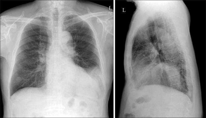
Figure 1 Chest radiographs show an increased opacity at the left lower lung field.

Figure 2 Chest CT scans reveal large inhomogeneous fat-containing mass in the left lower hemithorax with a mild enhancing soft tissue density portion. CT: computed tomography.
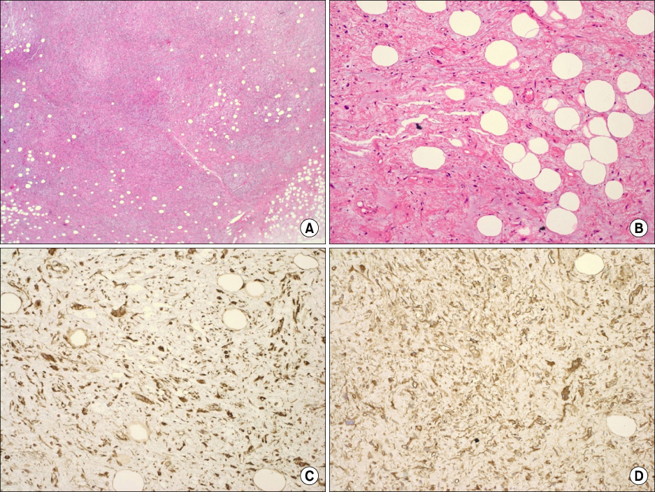
Figure 3 Percutaneous needle aspiration biopsy shows a mesenchymal neoplasm with spindle cells, a mature fat component and collagenous stroma suggestive of a solitary fibrous tumor, fat-containing variant (hematoxylin-eosin, original magnification ×100 (A), ×200 (B)). Immunostains show positive results for vimentin (C) and CD34 (D) (all original magnification, ×200).
Reference
-
1. Fletcher CD. The evolving classification of soft tissue tumours: an update based on the new WHO classification. Histopathology. 2006. 48:3–12.2. Lee JR, Kim JS, Lee CH, Sohn KR. A case of lipomatous hemangiopericytoma of the nasal cavity. Korean J Otolaryngol-Head Neck Surg. 2001. 44:893–896.3. Kim MY, Rha SE, Oh SN, Lee YJ, Byun JY, Jung CK, et al. Case report. Lipomatous haemangiopericytoma (fat-forming solitary fibrous tumour) involving the perineum: CT and MRI findings and pathological correlation. Br J Radiol. 2009. 82:e23–e26.4. Nielsen GP, Dickersin GR, Provenzal JM, Rosenberg AE. Lipomatous hemangiopericytoma. A histologic, ultrastructural and immunohistochemical study of a unique variant of hemangiopericytoma. Am J Surg Pathol. 1995. 19:748–756.5. Liu X, Zhang HY, Bu H, Meng GZ, Zhang Z, Ke Q. Fat-forming variant of solitary fibrous tumor of the mediastinum. Chin Med J (Engl). 2007. 120:1029–1032.6. Guillou L, Gebhard S, Coindre JM. Lipomatous hemangiopericytoma: a fat-containing variant of solitary fibrous tumor? Clinicopathologic, immunohistochemical, and ultrastructural analysis of a series in favor of a unifying concept. Hum Pathol. 2000. 31:1108–1115.7. Gengler C, Guillou L. Solitary fibrous tumour and haemangiopericytoma: evolution of a concept. Histopathology. 2006. 48:63–74.8. Cameselle-Teijeiro J, Manuel Lopes J, Villanueva JP, Gil-Gil P, Sobrinho-Simões M. Lipomatous haemangiopericytoma (adipocytic variant of solitary fibrous tumour) of the thyroid. Histopathology. 2003. 43:406–408.9. Pitchamuthu H, Gonzalez P, Kyle P, Roberts F. Fat-forming variant of solitary fibrous tumour of the orbit: the entity previously known as lipomatous haemangiopericytoma. Eye (Lond). 2009. 23:1479–1481.10. Robinson LA. Solitary fibrous tumor of the pleura. Cancer Control. 2006. 13:264–269.11. van de Rijn M, Lombard CM, Rouse RV. Expression of CD34 by solitary fibrous tumors of the pleura, mediastinum, and lung. Am J Surg Pathol. 1994. 18:814–820.12. Theunissen PH, Ariëns AT, Pannebakker MA, Blaauw G. Late recurrence of a hemangiopericytoma with lipomatous components. Pathologe. 1990. 11:346–349.13. Folpe AL, Devaney K, Weiss SW. Lipomatous hemangiopericytoma: a rare variant of hemangiopericytoma that may be confused with liposarcoma. Am J Surg Pathol. 1999. 23:1201–1207.14. Ceballos KM, Munk PL, Masri BA, O'Connell JX. Lipomatous hemangiopericytoma: a morphologically distinct soft tissue tumor. Arch Pathol Lab Med. 1999. 123:941–945.15. Domanski HA. Fine-needle aspiration smears from lipomatous hemangiopericytoma need not be confused with myxoid liposarcoma. Diagn Cytopathol. 2003. 29:287–291.
- Full Text Links
-
- Actions
-
Cited
- CITED
-
- Close
- Share
-
- Similar articles
-
- A Rare Solitary Fibrous Tumor of the Pleura with Extensive Cystic Change
- Ultrasonographic Localization of Solitary Fibrous Tumor of Pleura: Case Report
- A Case of Solitary Fibrous Tumor in the Cheek
- Pleural Localized Malignant Mesothelioma Mimicking a Benign Solitary Fibrous Tumor of the Pleura on Chest Computed Tomography: A Case Report
- Malignant Solitary Fibrous Tumor in Anterior Mediastinum with Pleural Metastasis Simulating Invasive Thymoma
