Clinical Benefits of Narrow Band Imaging Bronchoscopy in Central Lung Cancer
- Affiliations
-
- 1Divison of Pulmonary and Critical Care Medicine, Asan Medical Center, University of Ulsan College of Medicine, Seoul, Korea. ccm@amc.seoul.kr
- 2Department of Pathology, Asan Medical Center, University of Ulsan College of Medicine, Seoul, Korea.
Abstract
- BACKGROUND
Lung cancer is usually diagnosed at an advanced stage, resulting in a poor prognosis. The detection of these lesions at an earlier stage would be a clear benefit to patients. However, it is extremely difficult to detect carcinomatous lesions in the bronchial mucosal sites during a routine bronchoscopy.
METHODS
This study employed a novel optical technique, known as narrowband imaging (NBI), which allows noninvasive visualization of the microvascular structure of an organ's surface using reflected light.
RESULTS
Narrow band imaging was performed on 10 patients who were radiologically suspicious or had a high risk of lung cancer. The median age of the patients was 57.5 years (range, 44~81 years), and 80% of the patients were male. All lesions showed a microvascular proliferation pattern (dotted, tortuous and abruptly ending vessel) on the magnified NBI. Two lesions were confirmed histologically to be adenocarcinoma and the remaining lesions were squamous cell carcinomas. Two lesions were confirmed histologically to be a carcinoma in situ.
CONCLUSION
NBI is a promising and potentially powerful tool for identifying carcinomas at an earlier stage or a central lesion during a routine bronchoscopy examination.
Keyword
MeSH Terms
Figure
-
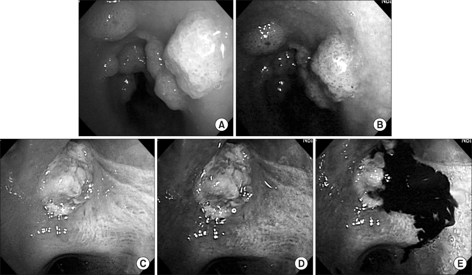
Figure 1 Multiple elevated nodular lesions in trachea were seen under white light (A) and narrow band imaging (B), more clearly visible with dotted vascularity in the narrow band imaging. Fungating mass without invasion in right upper lobe bronchus was seen under white light (C) and narrow band imaging (D). Peripheral margins of the abnormal neoangiogenetic vascularization are clearly visible and located well beyond the limits of the lesion as identified by white light bronchoscopy. It was easy to distinguish bleeding to neoangiogenetic vascularization under narrow band image (E).
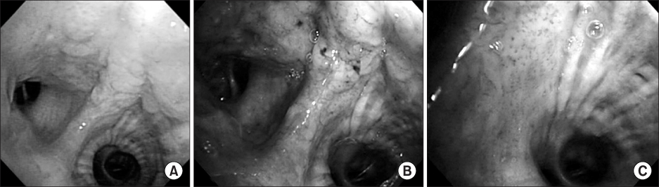
Figure 2 Mucosal irregularity in the superior segment of left lower lobe bronchus was seen under white light (A) and narrow band imaging (B). Dotted lesion under narrow band imaging was seen in right side of spur (C). Biopsy was performed at 3 sites (left, spur, right). Only dotted lesion on right revealed the squamous cell carcinoma. Other site was normal.
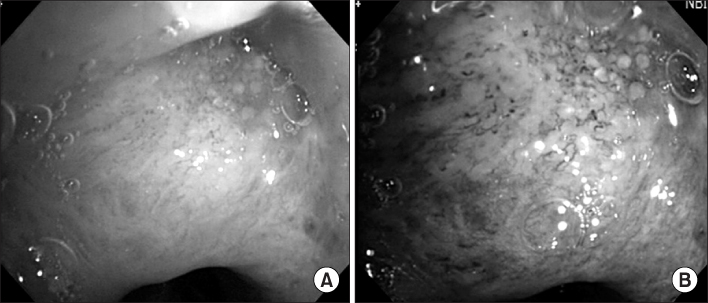
Figure 3 Tortous vascular lesion with abrupt ending was seen under white light (A) and narrow band imaging (B).
Reference
-
1. Shin HR, Won YJ, Jung KW, Park JG, Ahn YO. Cancer registration and statistics in Korea. J Korean Assoc Cancer Prev. 2004. 9:49–55.2. Korea National Statistical Office. Annual report on the cause of death statistics, 2006. 2007. Daejeon: Korea National Statistical Office;20–21.3. Humphrey LL, Teutsch S, Johnson M. Lung cancer screening with sputum cytologic examination, chest radiography, and computed tomography: an update for the U.S. Preventive Services Task Force. Ann Intern Med. 2004. 140:740–753.4. Infante M, Cavuto S, Lutman FR, Brambilla G, Chiesa G, Ceresoli G, et al. A randomized study of lung cancer screening with spiral computed tomography: three-year results from the DANTE trial. Am J Respir Crit Care Med. 2009. 180:445–453.5. Lam S, Becker HD. Future diagnostic procedures. Chest Surg Clin N Am. 1996. 6:363–380.6. Kim JO. Lung cancer screening. Tuberc Respir Dis. 2006. 61:207–213.7. Shibuya K, Hoshino H, Chiyo M, Iyoda A, Yoshida S, Sekine Y, et al. High magnification bronchovideoscopy combined with narrow band imaging could detect capillary loops of angiogenic squamous dysplasia in heavy smokers at high risk for lung cancer. Thorax. 2003. 58:989–995.8. Kim JO, Lee TH. The current status of virtual chromoscopy. Korean J Gastrointest Endosc. 2009. 38:309–322.9. Piazza C, Dessouky O, Peretti G, Cocco D, De Benedetto L, Nicolai P. Narrow-band imaging: a new tool for evaluation of head and neck squamous cell carcinomas: review of the literature. Acta Otorhinolaryngol Ital. 2008. 28:49–54.10. Kanao H, Tanaka S, Oka S, Hirata M, Yoshida S, Chayama K. Narrow-band imaging magnification predicts the histology and invasion depth of colorectal tumors. Gastrointest Endosc. 2009. 69(3 Pt 2):631–636.11. Zaric B, Becker HD, Perin B, Stojanovic G, Jovelic A, Eri Z, et al. Autofluorescence imaging videobrochoscopy improves assessment of tumor margins and affects therapeutic strategy in central lung cancer. Jpn J Clin Oncol. 2009. doi:10.1093/jjco/hyp135.12. Edell E, Lam S, Pass H, Miller YE, Sutedja T, Kennedy T, et al. Detection and localization of intraepithelial neoplasia and invasive carcinoma using fluorescence-reflectance bronchoscopy: an international, multicenter clinical trial. J Thorac Oncol. 2009. 4:49–54.13. Haussinger K, Becker H, Stanzel F, Kreuzer A, Schmidt B, Strausz J, et al. Autofluorescence bronchoscopy with white light bronchoscopy compared with white light bronchoscopy alone for the detection of precancerous lesions: a European randomised controlled multicentre trial. Thorax. 2005. 60:496–503.14. East JE, Tan EK, Bergman JJ, Saunders BP, Tekkis PP. Meta-analysis: narrow band imaging for lesion characterization in the colon, oesophagus, duodenal ampulla and lung. Aliment Pharmacol Ther. 2008. 28:854–867.15. Bojan Z, Branislav P, Aleksandra J, Goran S, Miroslav ID, Ilija A, et al. Influence of narrow band imaging (NBI) videobronchoscopy on the assessment of central lung cancer extension and therapeutic decision. Cancer Invest. 2009. 27:918–923.16. Goda K, Tajiri H, Ikegami M, Urashima M, Nakayoshi T, Kaise M. Usefulness of magnifying endoscopy with narrow band imaging for the detection of specialized intestinal metaplasia in columnar-lined esophagus and Barrett's adenocarcinoma. Gastrointest Endosc. 2007. 65:36–46.17. Herth FJ, Eberhardt R, Anantham D, Gompelmann D, Zakaria MW, Ernst A. Narrow-band imaging bronchoscopy increases the specificity of bronchoscopic early lung cancer detection. J Thorac Oncol. 2009. 4:1060–1065.18. Zaric B, Becker HD, Perin B, Jovelic A, Stojanovic G, Ilic MD, et al. Narrow band imaging videobronchoscopy improves assessment of lung cancer extension and influences therapeutic strategy. Jpn J Clin Oncol. 2009. 39:657–663.19. Konerding MA, Fait E, Gaumann A. 3D microvascular architecture of pre-cancerous lesions and invasive carcinomas of the colon. Br J Cancer. 2001. 84:1354–1362.20. Kumagai Y, Inoue H, Nagai K, Kawano T, Iwai T. Magnifying endoscopy, stereoscopic microscopy, and the microvascular architecture of superficial esophageal carcinoma. Endoscopy. 2002. 34:369–375.
- Full Text Links
-
- Actions
-
Cited
- CITED
-
- Close
- Share
-
- Similar articles
-
- Interobserver Agreement in Using Magnifying Narrow Band Imaging System
- A case of early-detected synchronous lung cancer by narrow-band imaging treated with photodynamic therapy
- Clinical Role of Magnifying Endoscopy with Narrow-band Imaging in the Diagnosis of Early Gastric Cancer
- Usefulness of Narrow-Band Imaging in Endoscopic Submucosal Dissection of the Stomach
- Application of artificial intelligence for diagnosis of early gastric cancer based on magnifying endoscopy with narrow-band imaging
