A Case of Bronchoscopic Treatment of a Bronchopleural Fistula Accompanied by Pneumonia
- Affiliations
-
- 1Department of Radiology, Donghae Hospital, Workers Accident Medical Coopration, Donghae, Korea. fovos@unitel.co.kr
Abstract
- A bronchopleural fistula (BPF) is traditionally treated by surgery, but currently various noninvasive forms of management, particularly the use of bronchoscopy, have been utilized. The substances and methods for noninvasive management of a BPF differ with individual clinicians. This case describes the use of flexible bronchoscopic treatment of a BPF complicating pneumoniausing embolization coils and intraluminally injected fibrin glue. If the BPF is small and is located on the peripheral bronchus, this minimal invasive maneuver could be recommended for the treatment of a BPF.
Keyword
Figure
-
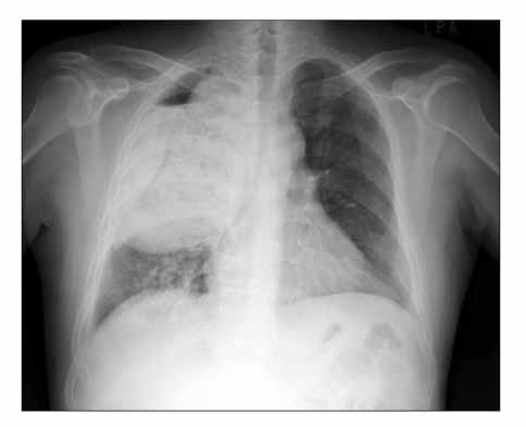
Figure 1 Initial chest X-ray finding of the patient: necrotizing pneumonia accompanying bronchopleural fistula in right upper lobe.
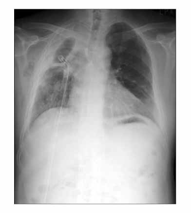
Figure 2 At three weeks after systemic antibiotics and closed thoracostomy for the treatment of bronchopleural fistula and necrotizing pneumonia, chest X-ray showed persistent dead space of right upper lobe. Also, minimal subcutaneous emphysema was noted in right axillary area.

Figure 3 Intraoperative X-ray finding during bronchoscopic coiling for treatment of bronchopleural fistula: there was no gastrograffin leakage into diseased bronchopleural tract after coiling and injection of glue. Before procedures, gastrograffin was used for localization of bronchopleural fistula tract, so small amount of leaked gastrograffin in both the dead space and chest tube is visible.
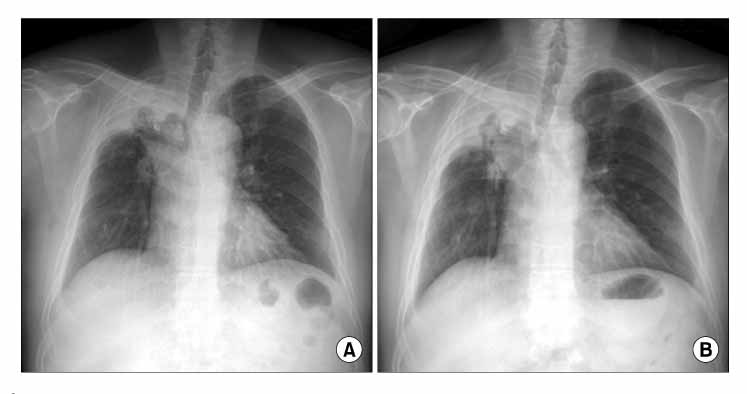
Figure 4 (A) Chest X-ray at discharge showed inserted colis into bronchopleural fistula and small amount of dead space and moderate pleural thickenings. (B) At 9 months postoperatively, chest x-ray revealed decreased pleural thickenings and well anchored coils in the bronchopleural fistula tract.
Reference
-
1. Puskas JD, Mathisen DJ, Grillo HC, Wain JC, Wright CD, Moncure AC. Treatment strategies for bronchopleural fistula. J Thorac Cardiovasc Surg. 1995. 109:989–995.2. Lois M, Noppen M. Bronchopleural fistulas: an overview of the problem with special focus on endoscopic management. Chest. 2005. 128:3955–3965.3. Galetta D, Veronesi G, Solli P, Petrella F, Borri A, Gasparri R, et al. A safe and effective method for an immediate bronchopleural fistula repair. Minerva Chir. 2007. 62:137–139.4. Hartmann W, Rausch V. New therapeutic application of the fiberoptic bronchoscope. Chest. 1997. 71:237.5. Na KJ, Kim BP, Hong SB, Choi YS, Kim SH, Ahn BH. Endobronchial closure of postoperative bronchopleural fistula using vascular occluding coils. Korean J Thorac Cardiovasc Surg. 2005. 38:72–75.6. Choi YI, Cho JH, Shim JY, Sheen SS, Oh YJ, Park JH, et al. A case of RUL bronchopleural fistula occluded by flexible bronchoscope with Endobronchial Watanabe Spigot (EWS). Tuberc Respir Dis. 2005. 58:404–409.7. Varoli F, Roviaro G, Grignani F, Vergani C, Maciocco M, Rebuffat C. Endoscopic treatment of bronchopleural fistulas. Ann Thorac Surg. 1998. 65:807–809.
- Full Text Links
-
- Actions
-
Cited
- CITED
-
- Close
- Share
-
- Similar articles
-
- Bronchoscopic Treatment of a Bronchopleural Fistula with using Coils and Fibrin Glue: A case report
- Use of the Free Flap for Large Defect with Bronchopleural Fistula: Case Report
- Closure of a Postoperative Bronchopleural Fistula with Bronchoscopic Instillation of n-butyl-2-cyanoacrylate (Histoacryl(R))
- A Case of Peripheral Bronchopleural Fistula Treated by Flexible Bronchoscopy with Gelfoam Occlusion
- Peripheral Bronchopleural Fistula: CT Evaluation in 22 patients
