J Korean Ophthalmol Soc.
2016 Jul;57(7):1102-1108. 10.3341/jkos.2016.57.7.1102.
Association between Metabolic Syndrome and Retinal Vascular Changes in Koreans based on Health Check-ups
- Affiliations
-
- 1Department of Ophthalmology, Inje University Sanggye Paik Hospital, Seoul, Korea. 991027js@hanmail.net
- KMID: 2317571
- DOI: http://doi.org/10.3341/jkos.2016.57.7.1102
Abstract
- PURPOSE
To evaluate the associations between components of metabolic syndrome and retinal vascular changes in a Korean population based on data collected at health check-ups.
METHODS
Fundus photographs of 381 patients participating in a health check-up were examined to identify central retinal artery equivalent (CRAE), central retinal vein equivalent (CRVE), and arteriovenous ratio (AVR) by IVAN software. Retinal hemorrhage, arteriovenous nicking, and retinal exudate were also noted. The association between metabolic syndrome and each component was then analyzed.
RESULTS
Significant associations were shown between metabolic syndrome and CRAE (p = 0.032), central obesity and CRAE (p = 0.037), triglyceride and CRAE (p = 0.011), and triglyceride and AVR (p = 0.005), in addition to central obesity and arteriovenous nicking (odds ratio [OR] = 2.68, p = 0.013), central obesity and retinal exudate (OR = 2.30, p = 0.038), serum glucose and retinal hemorrhage (OR = 8.06, p = 0.030), and blood pressure and arteriovenous nicking (OR = 2.78, p = 0.007).
CONCLUSIONS
Metabolic syndrome showed a significant relationship with retinal artery diameter. Central obesity showed the greatest relationship with retinal vascular changes among each of the components of metabolic syndrome.
MeSH Terms
Figure
-
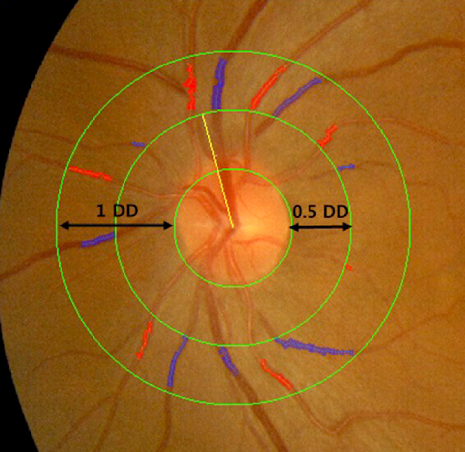
Figure 1. Fundus photograph centered at the optic disc of left eye, grader analysis measures arteries and veins, the largest six arteries (red) and veins (blue) are used to calculate the central retinal artery equivalent and central retinal vein equivalent by the “Big 6” method. Yellow line is the vessel indicator of the IVAN. DD = disc diameter.
Reference
-
References
1. Timar O, Sestier F, Levy E. Metabolic syndrome X: a review. Can J Cardiol. 2000; 16:779–89.2. National Cholesterol Education Program (NCEP) Expert Panel on Detection, Evaluation, and Treatment of High Blood Cholesterol in Adults (Adult Treatment Panel III). Third report of the national cholesterol education program (NCEP) expert panel on detection, evaluation, and treatment of high blood cholesterol in adults (adult treatment panel III) final report. Circulation. 2002; 106:3143–421.3. Grundy SM, Brewer HB Jr, Cleeman JI, et al. Definition of abdominal syndrome: report of the National Heart, Lung, and Blood Institute/American Heart Association conference on scientific abdominal related to definition. Circulation. 2004; 109:433–8.4. Ford ES, Giles WH, Dietz WH. Prevalence of the metabolic abdominal among US adults: findings from the third National Health and Nutrition Examination Survey. JAMA. 2002; 287:356–9.5. Park JS, Park HD, Yun JW, et al. Prevalence of the metabolic abdominal as defined by NCEP – ATPⅢ among the urban Korean population. Korean J Med. 2002; 63:290–8.6. Klein BE, Klein R, Lee KE. Components of the metabolic abdominal and risk of cardiovascular disease and diabetes in Beaver Dam. Diabetes Care. 2002; 25:1790–4.7. Golden SH, Folsom AR, Coresh J, et al. Risk factor groupings abdominal to insulin resistance and their synergistic effects on abdominal atherosclerosis: the atherosclerosis risk in communities study. Diabetes. 2002; 51:3069–76.8. de Jongh RT, Serné EH, IJzerman RG, et al. Impaired abdominal function in obesity: implications for obesity-associated microangiopathy, hypertension, and insulin resistance. Circulation. 2004; 109:2529–35.9. Wong TY, Kamineni A, Klein R, et al. Quantitative retinal venular caliber and risk of cardiovascular disease in older persons: the abdominal health study. Arch Intern Med. 2006; 166:2388–94.10. Ikram MK, de Jong FJ, Vingerling JR, et al. Are retinal arteriolar or venular diameters associated with markers for cardiovascular abdominal? The Rotterdam Study. Invest Ophthalmol Vis Sci. 2004; 45:2129–34.11. Wong TY, Islam FM, Klein R, et al. Retinal vascular caliber, abdominal risk factors, and inflammation: the abdominal study of atherosclerosis (MESA). Invest Ophthalmol Vis Sci. 2006; 47:2341–50.12. McGeechan K, Liew G, Macaskill P, et al. Meta-analysis: retinal vessel caliber and risk for coronary heart disease. Ann Intern Med. 2009; 151:404–13.
Article13. Ikram MK, de Jong FJ, Bos MJ, et al. Retinal vessel diameters and risk of stroke: the Rotterdam Study. Neurology. 2006; 66:1339–43.
Article14. McGeechan K, Liew G, Macaskill P, et al. Prediction of incident stroke events based on retinal vessel caliber: a systematic review and individual-participant meta-analysis. Am J Epidemiol. 2009; 170:1323–32.
Article15. Jeganathan VS, Sabanayagam C, Tai ES, et al. Retinal vascular abdominal and diabetes in a multiethnic Asian population. Microcirculation. 2009; 16:534–43.16. Islam FM, Nguyen TT, Wang JJ, et al. Quantitative retinal vascular calibre changes in diabetes and retinopathy: the Singapore Malay eye study. Eye (Lond). 2009; 23:1719–24.
Article17. Ikram MK, Janssen JA, Roos AM, et al. Retinal vessel diameters and risk of impaired fasting glucose or diabetes: the Rotterdam study. Diabetes. 2006; 55:506–10.18. Wong TY, Duncan BB, Golden SH, et al. Associations between the metabolic syndrome and retinal microvascular signs: the Atherosclerosis Risk In Communities study. Invest Ophthalmol Vis Sci. 2004; 45:2949–54.
Article19. Liew G, Sharrett AR, Wang JJ, et al. Relative importance of abdominalic determinants of retinal arteriolar and venular caliber: the atherosclerosis risk in communities study. Arch Ophthalmol. 2008; 126:1404–10.20. Klein R, Klein BE, Knudtson MD, et al. Are inflammatory factors related to retinal vessel caliber? The Beaver Dam Eye Study. Arch Ophthalmol. 2006; 124:87–94.21. Nguyen TT, Wang JJ, Sharrett AR, et al. Relationship of retinal vascular caliber with diabetes and retinopathy: the MultiEthnic Study of Atherosclerosis (MESA). Diabetes Care. 2008; 31:544–9.22. Kawasaki R, Tielsch JM, Wang JJ, et al. The metabolic syndrome and retinal microvascular signs in a Japanese population: the Funagata study. Br J Ophthalmol. 2008; 92:161–6.
Article23. Yuan Y, Ikram MK, Vingerling JR, et al. Retinal vascular caliber and metabolic syndrome in a Chinese population. Intern Med J. 2012; 42:1014–22.
Article24. Leung H, Wang JJ, Rochtchina E, et al. Computer-assisted retinal vessel measurement in an older population: correlation between right and left eyes. Clin Experiment Ophthalmol. 2003; 31:326–30.
Article25. Hubbard LD, Brothers RJ, King WN, et al. Methods for evaluation of retinal microvascular abnormalities associated with abdominal/sclerosis in the Atherosclerosis Risk in Communities Study. Ophthalmology. 1999; 106:2269–80.26. Knudtson MD, Lee KE, Hubbard LD, et al. Revised formulas for summarizing retinal vessel diameters. Curr Eye Res. 2003; 27:143–9.
Article27. Frisbee JC. Remodeling of the skeletal muscle microcirculation abdominals resistance to perfusion in obese Zucker rats. Am J Physiol Heart Circ Physiol. 2003; 285:H104–11.28. Oren S, Grossman E, Frohlich ED. Arterial and venous compliance in obese and nonobese subjects. Am J Cardiol. 1996; 77:665–7.
Article29. Klein R, Klein BE, Moss SE, et al. Retinal vascular caliber in abdominal with type 2 diabetes: the Wisconsin Epidemiological Study of Diabetic Retinopathy: XX. Ophthalmology. 2006; 113:1488–98.30. Tikellis G, Wang JJ, Tapp R, et al. The relationship of retinal abdominal calibre to diabetes and retinopathy: the Australian Diabetes, Obesity and Lifestyle (AusDiab) study. Diabetologia. 2007; 50:2263–71.31. Curtis TM, Gardiner TA, Stitt AW. Microvascular lesions of abdominal retinopathy: clues towards understanding pathogenesis? Eye (Lond). 2009; 23:1496–508.32. Kawasaki R, Cheung N, Wang JJ, et al. Retinal vessel diameters and risk of hypertension: the Multiethnic Study of Atherosclerosis. J Hypertens. 2009; 27:2386–93.
Article33. Cheung N, Sharrett AR, Klein R, et al. Aortic distensibility and abdominal arteriolar narrowing: the abdominal study of atherosclerosis. Hypertension. 2007; 50:617–22.34. Hemminki V, Kähönen M, Tuomisto MT, et al. Determination of retinal blood vessel diameters and arteriovenous ratios in systemic hypertension: comparison of different calculation formulae. Graefes Arch Clin Exp Ophthalmol. 2007; 245:8–17.
Article
- Full Text Links
-
- Actions
-
Cited
- CITED
-
- Close
- Share
-
- Similar articles
-
- Association of Participation in Health Check-ups with Risk Factors for Cardiovascular Diseases
- Efficacy of Retinal Check-up Using Fundus Photography in Patients with Epiretinal Membrane
- Association between Serum Carcinoembryonic Antigen Levels within Normal Range and Metabolic Syndrome in Korean Women Aged ≥50 Years Old
- Comparison between a Pediatric Health Promotion Center and a Pediatric Obesity Clinic in Detecting Metabolic Syndrome and Non-Alcoholic Fatty Liver Disease in Children
- Factors Related to Physical Health Monitoring in Community-Dwelling Patients with Schizophrenia Spectrum Disorder
