Restor Dent Endod.
2015 Feb;40(1):75-78. 10.5395/rde.2015.40.1.75.
Endodontic treatment of a mandibular first molar with 8 canals: a case report
- Affiliations
-
- 1Department of Conservative Dentistry and Endodontics, M.P. Dental College, Hospital and Oral Research Institute, Vadodara, India. aroraankit24@gmail.com
- 2Department of Conservative Dentistry and Endodontics, Manipal College of Dental Sciences, Manipal University, Karnataka, India.
- 3Department of Orthodontics and Dentofacial Orthopaedics, M.P. Dental College, Hospital and Oral Research Institute, Vadodara, India.
- KMID: 2316927
- DOI: http://doi.org/10.5395/rde.2015.40.1.75
Abstract
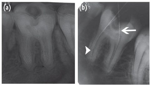
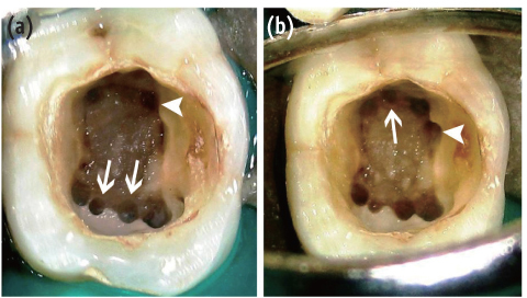
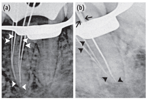
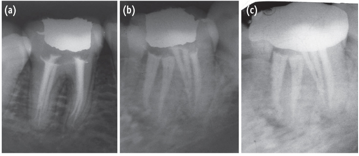
- Presented here is a case where 8 canals were located in a mandibular first molar. A patient with continuing pain in mandibular left first molar even after completion of biomechanical preparation was referred by a dentist. Following basic laws of the pulp chamber floor anatomy, 8 canals were located in three steps with 4 canals in each root. In both of the roots, 4 separate canals commenced which joined into two canals and exited as two separate foramina. At 6 mon follow-up visit, the tooth was found to be asymptomatic and revealed normal radiographic periapical area. The case stresses on the fact that understanding the laws of pulp chamber anatomy and complying with them while attempting to locate additional canals can prevent missing canals.
Figure
Reference
-
1. Vertucci FJ. Root canal anatomy of the human permanent teeth. Oral Surg Oral Med Oral Pathol. 1984; 58:589–599.
Article2. Vertucci FJ. Root canal morphology and its relationship to endodontic procedures. Endod Topics. 2005; 10:3–29.
Article3. Cantatore G, Berutti E, Castellucci A. Missed anatomy: frequency and clinical impact. Endod Topics. 2006; 15:3–31.
Article4. Krasner P, Rankow HJ. Anatomy of the pulp-chamber floor. J Endod. 2004; 30:5–16.
Article5. Carr GB, Murgel CA. The use of the operating microscope in endodontics. Dent Clin North Am. 2010; 54:191–214.
Article6. Cotton TP, Geisler TM, Holden DT, Schwartz SA, Schindler WG. Endodontic applications of cone-beam volumetric tomography. J Endod. 2007; 33:1121–1132.
Article7. de Pablo OV, Estevez R, Péix Sánchez M, Heilborn C, Cohenca N. Root anatomy and canal configuration of the permanent mandibular first molar: a systematic review. J Endod. 2010; 36:1919–1931.
Article8. Nur BG, Ok E, Altunsoy M, Aglarci OS, Colak M, Gungor E. Evaluation of the root and canal morphology of mandibular permanent molars in a south-eastern Turkish population using cone-beam computed tomography. Eur J Dent. 2014; 8:154–159.
Article9. Martinez-Berna A, Badanelli P. Mandibular first molars with six root canals. J Endod. 1985; 11:348–352.
Article10. Reeh ES. Seven canals in a lower first molar. J Endod. 1998; 24:497–499.
Article11. Mortman RE, Ahn S. Mandibular first molars with three mesial canals. Gen Dent. 2003; 51:549–551.12. Baziar H, Daneshvar F, Mohammadi A, Jafarzadeh H. Endodontic management of a mandibular first molar with four canals in a distal root by using cone-beam computed tomography: a case report. J Oral Maxillofac Res. 2014; 04. 01. 5(1):e5. doi: 10.5037/jomr.2014.5105. [Epub ahead of print].
Article13. Valerian Albuquerque D, Kottoor J, Velmurugan N. A new anatomically based nomenclature for the roots and root canals-part 2: mandibular molars. Int J Dent. 2012; 2012:814789. doi: 10.1155/2012/814789. [Epub ahead of print].
Article14. Kottoor J, Sudha R, Velmurugan N. Middle distal canal of the mandibular first molar: a case report and literature review. Int Endod J. 2010; 43:714–722.
Article15. Baugh D, Wallace J. Middle mesial canal of the mandibular first molar: a case report and literature review. J Endod. 2004; 30:185–186.
Article16. Faramarzi F, Fakhri H, Javaheri HH. Endodontic treatment of a mandibular first molar with three mesial canals and broken instrument removal. Aust Endod J. 2010; 36:39–41.
Article17. Goel NK, Gill KS, Taneja JR. Study of root canals configuration in mandibular first permanent molar. J Indian Soc Pedod Prev Dent. 1991; 8:12–14.18. Jacobsen EL, Dick K, Bodell R. Mandibular first molars with multiple mesial canals. J Endod. 1994; 20:610–613.
Article19. Kontakiotis EG, Tzanetakis GN. Four canals in the mesial root of a mandibular first molar. A case report under the operating microscope. Aust Endod J. 2007; 33:84–88.
Article20. Aminsobhani M, Shokouhinejad N, Ghabraei S, Bolhari B, Ghorbanzadeh A. Retreatment of a 6-canalled mandibular frirst molar with four mesial canals: a case report. Iran Endod J. 2010; 5:138–140.21. al-Nazhan S. Incidence of four canals in root-canal-treated mandibular first molars in a Saudi Arabian subpopulation. Int Endod J. 1999; 32:49–52.
Article22. Beatty RG, Interian CM. A mandibular first molar with five canals: report of case. J Am Dent Assoc. 1985; 111:769–771.
Article23. Friedman S, Moshonov J, Stabholz A. Five root canals in a mandibular first molar. Endod Dent Traumatol. 1986; 2:226–228.
Article24. Gulabivala K, Aung TH, Alavi A, Ng YL. Root and canal morphology of Burmese mandibular molars. Int Endod J. 2001; 34:359–370.
Article25. Peiris HR, Pitakotuwage TN, Takahashi M, Sasaki K, Kanazawa E. Root canal morphology of mandibular permanent molars at different ages. Int Endod J. 2008; 41:828–835.
Article26. Nosonowitz DM, Brenner MR. The major canals of the mesiobuccal root of the maxillary 1st and 2nd molars. N Y J Dent. 1973; 43:12–15.27. Seidberg BH, Altman M, Guttuso J, Suson M. Frequency of two mesiobuccal root canals in maxillary permanent first molars. J Am Dent Assoc. 1973; 87:852–856.
Article
- Full Text Links
- Actions
-
Cited
- CITED
-
- Close
- Share
- Similar articles
-
- Endodontic treatment of a C-shaped mandibular second premolar with four root canals and three apical foramina: a case report
- A retrospective study on incidence of C-shaped canals in mandibular second molars
- Healing outcomes of root canal treatment for C-shaped mandibular second molars: a retrospective analysis
- Management of a permanent maxillary first molar with unusual crown and root anatomy: a case report
- An evaluation of canal curvature at the apical one third in type II mesial canals of mandibular molars


