Restor Dent Endod.
2014 Aug;39(3):180-186.
Enamel pretreatment with Er:YAG laser: effects on the microleakage of fissure sealant in fluorosed teeth
- Affiliations
-
- 1Department of Pediatric Dentistry, School of Dentistry, Shiraz University of Medical Sciences, Shiraz, Iran. memarpour@sums.ac.ir
- 2Private Dentist, Shiraz, Iran.
Abstract
OBJECTIVES
The purpose of this in vitro study was to evaluate the microleakage and penetration of fissure sealant in permanent molar teeth with fluorosis after pretreatment of the occlusal surface.
MATERIALS AND METHODS
A total of 120 third molars with mild dental fluorosis were randomly divided into 6 groups (n = 20). The tooth surfaces were sealed with an unfilled resin fissure sealant (FS) material. The experimental groups included: 1) phosphoric acid etching (AE) + FS (control); 2) AE + One-Step Plus (OS, Bisco) + FS; 3) bur + AE + FS; 4) bur + AE + OS + FS; 5) Er:YAG laser + AE + FS; and 6) Er:YAG laser + AE + OS + FS. After thermocycling, the teeth were immersed in 0.5% fuchsin and sectioned. Proportions of mircoleakage (PM) and unfilled area (PUA) were measured by digital microscope.
RESULTS
Overall, there were significant differences among all groups in the PM (p = 0.00). Group 3 showed the greatest PM, and was significantly different from groups 2 to 6 (p < 0.05). Group 6 showed the lowest PM. Pretreatment with Er:YAG with or without adhesive led to less PM than bur pretreatment. There were no significant differences among groups in PUA.
CONCLUSIONS
Conventional acid etching provided a similar degree of occlusal seal in teeth with fluorosis compared to those pretreated with a bur or Er:YAG laser. Pretreatment of pits and fissures with Er:YAG in teeth with fluorosis may be an alternative method before fissure sealant application.
Keyword
MeSH Terms
Figure
-
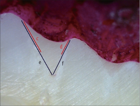
Figure 1 Method used to calculate the proportion of microleakage: (c + d) / (e + f). c and d represent microleakage in mm, and e and f represent the total length of fissure sealant in mm.21,28
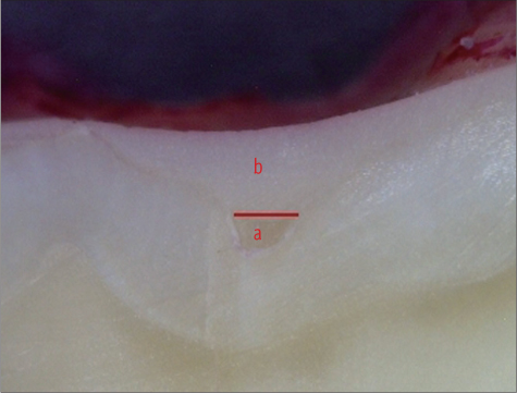
Figure 2 Method used to calculate the proportion of the unfilled area: a / (a + b). b represents the fissure sealant area and a represents the unfilled area.21,28
Reference
-
1. Beauchamp J, Caufield PW, Crall JJ, Donly K, Feigal R, Gooch B, Ismail A, Kohn W, Siegal M, Simonsen R. American Dental Association Council on Scientific Affairs. Evidence-based clinical recommendations for the use of pit-and-fissure sealants: a report of the American Dental Association Council on Scientific Affairs. J Am Dent Assoc. 2008; 139:257–268.
Article2. Simonsen RJ. Pit and fissure sealant: review of the literature. Pediatr Dent. 2002; 24:393–414.3. Sasa I, Donly KJ. Sealants: a review of the materials and utilization. J Calif Dent Assoc. 2010; 38:730–734.4. Cehreli ZC, Gungor HC. Quantitative microleakage evaluation of fissure sealants applied with or without a bonding agent: results after four-year water storage in vitro. J Adhes Dent. 2008; 10:379–384.5. Subramaniam P, Babu KL, Naveen HK. Effect of tooth preparation on sealant success-an in vitro study. J Clin Pediatr Dent. 2009; 33:325–331.6. Chaitra TR, Subba RV, Devarasa GM, Ravishankar TL. Microleakage and SEM analysis of flowable resin used as a sealant following three fissure preparation techniques-an in vitro study. J Clin Pediatr Dent. 2011; 35:277–282.
Article7. Geiger SB, Gulayev S, Weiss EI. Improving fissure sealant quality: mechanical preparation and filling level. J Dent. 2000; 28:407–412.
Article8. Lupi-Pégurier L, Muller-Bolla M, Bertrand MF, Fradet T, Bolla M. Microleakage of a pit-and-fissure sealant: effect of air-abrasion compared with classical enamel preparations. J Adhes Dent. 2004; 6:43–48.9. Agrawal A, Shigli A. Comparison of six different methods of cleaning and preparing occlusal fissure surface before placement of pit and fissure sealant: an in vitro study. J Indian Soc Pedod Prev Dent. 2012; 30:51–55.
Article10. Mazzoleni S, De Francesco M, Perazzolo D, Favero L, Bressan E, Ferro R, Stellini E. Comparative evaluation of different techniques of surface preparation for occlusal sealing. Eur J Paediatr Dent. 2007; 8:119–123.11. Olivi G, Margolis F, Genovese MD. Chapter 3. Hard tissue laser applications. Pediatric laser dentistry. A user's guide. 1st ed. Hanover Park, IL: Quintessence Publishing Co, Inc.;2011. p. 83–85.12. American Academy of Pediatric Dentistry, Council on Clinical Affairs. Policy on the use of lasers for pediatric pental patients. updated 2013 May 7. Available from: http://www.aapd.org/media/Policies_Guidelines/P_LasersUse.pdf.13. Sancakli HS, Erdemir U, Yildiz E. Effects of Er:YAG laser and air abrasion on the microleakage of a resin-based fissure sealant material. Photomed Laser Surg. 2011; 29:485–492.
Article14. Lasmar MF, Reher VG, Lalloo R, Reher P. Enamel demineralization and bracket bond strength when etching with acid and/or Er:YAG laser. Aust Dent J. 2012; 57:190–195.
Article15. Lupi-Pégurier L, Bertrand MF, Genovese O, Rocca JP, Muller-Bolla M. Microleakage of resin-based sealants after Er:YAG laser conditioning. Lasers Med Sci. 2007; 22:183–188.
Article16. Baygin O, Korkmaz FM, Tüzüner T, Tanriver M. The effect of different enamel surface treatments on the microleakage of fissure sealants. Lasers Med Sci. 2012; 27:153–160.
Article17. Borsatto MC, Corona SA, Ramos RP, Liporaci JL, Pécora JD, Palma-Dibb RG. Microleakage at sealant/enamel interface of primary teeth: effect of Er:YAG laser ablation of pits and fissures. J Dent Child (Chic). 2004; 71:143–147.18. Karaman E, Yazici AR, Baseren M, Gorucu J. Comparison of acid versus laser etching on the clinical performance of a fissure sealant: 24-month results. Oper Dent. 2013; 38:151–158.
Article19. Manhart J, Huth KC, Chen HY, Hickel R. Influence of the pretreatment of occlusal pits and fissures on the retention of a fissure sealant. Am J Dent. 2004; 17:12–18.20. Youssef MN, Youssef FA, Souza-Zaroni WC, Turbino ML, Vieira MM. Effect of enamel preparation method on in vitro marginal microleakage of a flowable composite used as pit and fissure sealant. Int J Paediatr Dent. 2006; 16:342–347.21. Khogli AE, Cauwels R, Vercruysse C, Verbeeck R, Martens L. Microleakage and penetration of a hydrophilic sealant and a conventional resin-based sealant as a function of preparation techniques: a laboratory study. Int J Paediatr Dent. 2013; 23:13–22.
Article22. Loyola-Rodriguez JP, Mendoza-Razo V, Rodriguez-Juarez F, Campos-Cambranis R. Flowable resin used as a sealant in molars affected by dental fluorosis: a comparative study. J Clin Pediatr Dent. 2005; 30:39–43.
Article23. Denbesten P, Li W. Chronic fluoride toxicity: dental fluorosis. Monogr Oral Sci. 2011; 22:81–96.
Article24. Torres-Gallegos I, Zavala-Alonso V, Patiño-Marín N, Martinez-Castañon GA, Anusavice K, Loyola-Rodríguez JP. Enamel roughness and depth profile after phosphoric acid etching of healthy and fluorotic enamel. Aust Dent J. 2012; 57:151–156.
Article25. Ateyah N, Akpata E. Factors affecting shear bond strength of composite resin to fluorosed human enamel. Oper Dent. 2000; 25:216–222.26. Al-Sugair MH, Akpata ES. Effect of fluorosis on etching of human enamel. J Oral Rehabil. 1999; 26:521–528.
Article27. Opinya GN, Pameijer CH. Tensile bond strength of fluorosed Kenyan teeth using the acid etch technique. Int Dent J. 1986; 36:225–229.28. Ciucchi P, Neuhaus KW, Emerich M, Peutzfeldt A, Lussi A. Evaluation of different types of enamel conditioning before application of a fissure sealant. Lasers Med Sci. 2013; 05. 01. [Epub ahead of print].
Article29. Hossain M, Yamada Y, Masuda-Murakami Y, Nakamura Y. Removal of organic debris with Er:YAG laser irradiation and microleakage of fissures sealants in vitro. Lasers Med Sci. 2012; 27:895–902.
Article30. Salama FS, Al-Hammad NS. Marginal seal of sealant and compomer materials with and without enameloplasty. Int J Paediatr Dent. 2002; 12:39–46.
Article31. Burrow JF, Burrow MF, Makinson OF. Pits and fissures: relative space contribution in fissures from sealants, prophylaxis pastes and organic remnants. Aust Dent J. 2003; 48:175–179.
Article32. Burrow MF, Burrow JF, Makinson OF. Pits and fissures: etch resistance in prismless enamel walls. Aust Dent J. 2001; 46:258–262.
Article33. Almerich-Silla JM, Montiel-Company JM, Ruiz-Miravet A. Caries and dental fluorosis in a western Saharan population of refugee children. Eur J Oral Sci. 2008; 116:512–517.
Article34. Blackwood JA, Dilley DC, Roberts MW, Swift EJ Jr. Evaluation of pumice, fissure enameloplasty and air abrasion on sealant microleakage. Pediatr Dent. 2002; 24:199–203.35. Walsh LJ. The current status of laser applications in dentistry. Aust Dent J. 2003; 48:146–155.
Article36. Moshonov J, Stabholz A, Zyskind D, Sharlin E, Peretz B. Acid-etched and erbium:yttrium aluminium garnet laser-treated enamel for fissure sealants: a comparison of microleakage. Int J Paediatr Dent. 2005; 15:205–209.
Article37. Son JH, Kim HC, Hur B, Park JK. The effect of Er,Cr: YSGG irradiation on microtensile bond strength of composite resin restoration. J Korean Acad Conserv Dent. 2010; 35:134–142.
Article38. Lygidakis NA, Dimou G, Stamataki E. Retention of fissure sealants using two different methods of application in teeth with hypomineralised molars (MIH): a 4 year clinical study. Eur Arch Paediatr Dent. 2009; 10:223–236.
Article39. Memarpour M, Shafiei F. Comparison of 3 one-bottle adhesives on fissure sealant microleakage: an in vitro study. J Dent Child (Chic). 2013; 80:16–19.40. Kim JH, Cho YG. Microshear bond strength of a flowable resin to enamel according to the different adhesive systems. J Korean Acad Conserv Dent. 2011; 36:50–58.
Article41. Kakaboura A, Matthaiou L, Papagiannoulis L. In vitro study of penetration of flowable resin composite and compomer into occlusal fissures. Eur J Paediatr Dent. 2002; 3:205–209.42. Kantovitz KR, Pascon FM, Alonso RC, Nobre-dos-Santos M, Rontani RM. Marginal adaptation of pit and fissure sealants after thermal and chemical stress. A SEM study. Am J Dent. 2008; 21:377–382.
- Full Text Links
-
- Actions
-
Cited
- CITED
-
- Close
- Share
-
- Similar articles
-
- Orthodontic bracket shear bond strength to ND:YAG laser and ER:YAG laser irradiated enamel
- Evaluation of different enamel conditioning techniques for orthodontic bonding
- The effects of a sealant resin on enamel demineralization in orthodontic bracket bonding
- A study on the enamel surface texture and caries susceptibility in interdentally stripped teeth
- Efficiency of ceramic bracket debonding with the Er:YAG laser
