Considerations during crown reattachment procedure over the pulpal exposure: case report
- Affiliations
-
- 1Department of Conservative Dentistry, Gangnam Severance Dental Hospital, Yonsei University College of Dentistry, Seoul, Korea. pjw@yuhs.ac
- 2Department of Conservative Dentistry, Yonsei University Wonju College of Medicine, Wonju, Korea.
Abstract
- Crown reattachment is the most conservative treatment which can be used to restore fractured tooth, presumably with sufficient strength, while maintaining original contour, incisal translucency, and reducing chair time and cost. However, in case of crown fracture with pin-point pulp exposure, we should cautiously minimize the irritation to the pulp and consider pre-treatment pulpal status, choice of pulp capping materials, choice of bonding system and treatment sequence during crown reattachment procedures. This case reports the considerations while crown reattachment with direct pulp capping using calcium hydroxide (Dycal, Dentsply Caulk).
Keyword
MeSH Terms
Figure
-

Figure 1 Initial visit. (a) Intraoral photograph (palatal view). Pin-point pulp exposure was seen on tooth #11 and 12; (b) Intraoral photograph (Labial view); (c) Periapical radiograph.
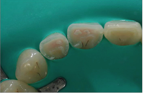
Figure 2 After acid etching, Dycal was applied.
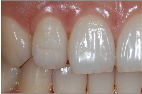
Figure 3 Labial view after the reattachment was done. The fracture line was visible, but the overcontouring was delayed to reduce the palpal damage.
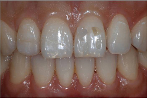
Figure 4 Shade selection was done from cervical and incisal area.
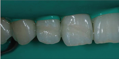
Figure 5 Fracture lines were prepared with 0.3 mm depth and 2.5 mm bevel in the incisal-cervical direction.
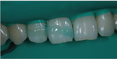
Figure 6 37% Phosphoric acid was applied to the fracture line for 30 seconds.
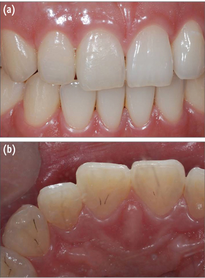
Figure 7 Composite was overcontoured and the excess was removed and polished. (a) Labial view; (b) Palatal view.

Figure 8 Immediate postoperative periapical view.

Figure 9 14 months after the treatment. (a) Labial view; (b) Palatal view; (c) Periapical view. The teeth had normal pulp and apex and was esthetically acceptable.
Reference
-
1. Ojeda-Gutierrez F, Martinez-Marquez B, Rosales-Ibanez R, Pozos-Guillen AJ. Reattachment of anterior teeth fragments using a modified Simonsen's technique after dental trauma: report of a case. Dent Traumatol. 2011. 27:81–85.
Article2. de Blanco LP. Treatment of crown fractures with pulp exposure. Oral Surg Oral Med Oral Pathol Oral Radiol Endod. 1996. 82:564–568.
Article3. Reis A, Kraul A, Francci C, de Assis TG, Crivelli DD, Oda M, Loguercio AD. Re-attachment of anterior fractured teeth: fracture strength using different materials. Oper Dent. 2002. 27:621–627.4. Hargreaves KM, Cohen SR. Cohen's pathways of the pulp expert consult. 2011. 10th ed. St. Louis: Mosby;627.5. Summitt JB, Robbins JW, Hilton T, Schwartz RS. Fundamentals of operative dentistry: a contemporary approach. 2006. 3rd ed. Hanover Park: Quintessence Publishing;220.6. Olsburgh S, Jacoby T, Krejci I. Crown fractures in the permanent dentition: pulpal and restorative considerations. Dent Traumatol. 2002. 18:103–115.
Article7. Liebenberg WH. Direct pulp capping considerations during tooth fragment reattachment: a case report. J Dent Assoc S Afr. 1993. 48:28–32.8. Macedo GV, Ritter AV. Essentials of rebonding tooth fragments for the best functional and esthetic outcomes. Pediatr Dent. 2009. 31:110–116.9. Pameijer CH, Stanley HR. The disastrous effects of the "total etch" technique in vital pulp capping in primates. Am J Dent. 1998. 11:S45–S54.10. Burke FJ, Watts DC. Weight loss of four calcium hydroxide-based materials following a phosphoric acid etching and washing cycle. J Dent. 1986. 14:226–227.
Article11. El-Araby A, Al-Jabab A. The influence of some dentin primers on calcium hydroxide lining cement. J Contemp Dent Pract. 2005. 6:1–9.
Article
- Full Text Links
-
- Actions
-
Cited
- CITED
-
- Close
- Share
-
- Similar articles
-
- Treatment of crown-root fracture with a modified crown fragment reattachment technique
- Considerations in the selection of method for clinical crown lengthening
- Reattachment of a fractured fragment with relined fiber post using indirect technique: a case report
- Conservative and esthetic approach in crown fracture of maxillay anterior tooth: tooth fragment reattachment
- Management of complicated crown fracture by tooth fragment reattachment with fiber post: a case report
