Alternative surgical approaches for aggressive angiomyxoma at different sites in the pelvic cavity
- Affiliations
-
- 1Department of Obstetrics and Gynecology, Inje University Haeundae Paik Hospital, Inje University College of Medicine, Busan, Korea. jyimdog@paik.ac.kr
- KMID: 2314072
- DOI: http://doi.org/10.5468/ogs.2015.58.6.525
Abstract
- Aggressive angiomyxoma, a rare soft tissue benign neoplasm, predominantly occurs in the female pelvic peritoneum and perineum region during reproductive age. It is slow growing, locally infiltrative, and has a high risk of local recurrence and the neoplastic character of blood vessels. The standard treatment is surgery. We report three unusual aggressive angiomyxoma cases. The first case was a pedunculated mass of the left labium major; the second, a left perineal mass that infiltrated into the paravesical area via the obturator foramen; and the third, a big mass in the retroperitoneal cavity, found that growing aggressive angiomyxoma looked like lava expulsion in the pelvic area. After a thorough examination and full radiologic workup, we performed surgical excision in each patient via different approaches. Histopathologic findings were consistent with diagnosis of aggressive angiomyxoma. To date, no relapse has been observed.
Keyword
MeSH Terms
Figure
-
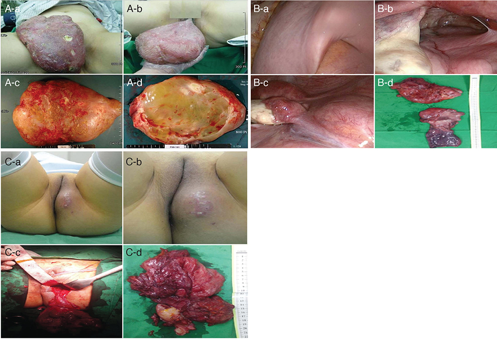
Fig. 1 Aggressive angiomyxoma: gross finding. (A) Case 1. A huge perineal mass that reveals aggressive angiomyxoma. (A-a,b) A huge polypoid pedunculated mass that measures 27 cm, with an attached stalk and overlying skin on the left labium is observed. (A-c,d) The mass shows a glistening surface and gelatinous cut surface. (B) Case 2. The retroperitoneal aggressive growth looks like lava expulsion. (B-a) The image shows the pelvic brim. (B-b) The image shows the left adnexal area and posterior cul-de-sac. (B-c) The image shows the protruding mass in the right pelvic retroperitoneum. (B-d) The image shows the excised tumor. (C) Case 3. The tumor lesion extended from the left paravesical space and obturator space, retroperitoneum to the left buttock. (C-a,b) External showing of mass on left labium major to left buttock. (C-c,d) Mass removed from retroperitoneal space which extended to paravesical and obtulator space, over 20-cm-sized irregular shaped.

Fig. 2 Radiologic image and microscopic finding. (A) A computed tomography scan in case 2 shows an 18×15×8-cm large, high-density cystic mass, suspected to be fluid collection in the pelvis, with diffuse peritoneal thickening. (A-a) Sagittal view and (A-b) coronal view. (B) This magnetic resonance imaging scan in case 3 shows a 15×10×6-cm large, irregularly shaped enhanced mass in the left perineum, extending to left retroperitoneum via the obturator foramen. (B-a) Coronal view of pelvis on T2 weighted image. (B-b) Sagittal view on T2 weighted image, distinguished with bladder, uterus, and rectum. (C) This image shows the microscopic photograph of the aggressive angiomyxoma (H&E stain, ×200).
Cited by 1 articles
-
Robotic extralevator excision of a retrorectal giant aggressive angiomyxoma
Scott R. Kelley
Obstet Gynecol Sci. 2018;61(6):693-697. doi: 10.5468/ogs.2018.61.6.693.
Reference
-
1. Kura MM, Jindal SR, Khemani UN. Aggressive angiomyxoma of the vulva: an uncommon entity. Indian Dermatol Online J. 2012; 3:128–130.2. Dahiya K, Jain S, Duhan N, Nanda S, Kundu P. Aggressive angiomyxoma of vulva and vagina: a series of three cases and review of literature. Arch Gynecol Obstet. 2011; 283:1145–1148.3. Sereda D, Sauthier P, Hadjeres R, Funaro D. Aggressive angiomyxoma of the vulva: a case report and review of the literature. J Low Genit Tract Dis. 2009; 13:46–50.4. Amin A, El Badawy S, Bull A. Aggressive angiomyxoma of the vulva. J Obstet Gynaecol. 2013; 33:325–326.5. Mandal S, Dhingra K, Roy S, Khurana N. Aggressive angiomyxoma of the vulva presenting as a pedunculated swelling. Indian J Pathol Microbiol. 2008; 51:259–260.6. Mathieson A, Chandrakanth S, Yousef G, Wadden P. Aggressive angiomyxoma of the pelvis: a case report. Can J Surg. 2007; 50:228–229.7. Dierickx I, Deraedt K, Poppe W, Verguts J. Aggressive angiomyxoma of the vulva: a case report and review of literature. Arch Gynecol Obstet. 2008; 277:483–487.8. Outwater EK, Marchetto BE, Wagner BJ, Siegelman ES. Aggressive angiomyxoma: findings on CT and MR imaging. AJR Am J Roentgenol. 1999; 172:435–438.9. Nyam DC, Pemberton JH. Large aggressive angiomyxoma of the perineum and pelvis: an alternative approach. Report of a case. Dis Colon Rectum. 1998; 41:514–516.10. Nucci MR, Fletcher CD. Vulvovaginal soft tissue tumours: update and review. Histopathology. 2000; 36:97–108.11. Basak S, Rogers S, Solomonsz AF. Superficial angiomyxoma of the vulva: a case report of a rare cutaneous tumour. J Obstet Gynaecol. 2011; 31:360–361.12. Sun NX, Li W. Aggressive angiomyxoma of the vulva: case report and literature review. J Int Med Res. 2010; 38:1547–1552.13. McCluggage WG, Jamieson T, Dobbs SP, Grey A. Aggressive angiomyxoma of the vulva: dramatic response to gonadotropin-releasing hormone agonist therapy. Gynecol Oncol. 2006; 100:623–625.14. Shinohara N, Nonomura K, Ishikawa S, Seki H, Koyanagi T. Medical management of recurrent aggressive angiomyxoma with gonadotropin-releasing hormone agonist. Int J Urol. 2004; 11:432–435.
- Full Text Links
-
- Actions
-
Cited
- CITED
-
- Close
- Share
-
- Similar articles
-
- Aggressive Angiomyxoma in Female Pelvic Cavity: A Case Report
- Aggressive Angiomyxoma at Ischiorectal Fossa
- Aggressive Angiomyxoma in the Vulva: A Case Report
- A case of recurrent aggressive angiomyxoma of the vulva in the adolescence
- Robotic extralevator excision of a retrorectal giant aggressive angiomyxoma
