Necrotizing ruptured vaginal leiomyoma mimicking a malignant neoplasm
- Affiliations
-
- 1Department of Obstetrics and Gynecology, Gangnam CHA Hospital, CHA University, Seoul, Korea.
- 2Department of Radiology, Gumi CHA Hospital, CHA University, Gumi, Korea.
- 3Department of Pathology, Gumi CHA Hospital, CHA University, Gumi, Korea.
- 4Department of Obstetrics and Gynecology, Gumi CHA Hospital, CHA University, Gumi, Korea. shsong8@gmail.com
- KMID: 2314029
- DOI: http://doi.org/10.5468/ogs.2014.57.6.560
Abstract
- Leiomyomas are common benign uterine tumors. However, the incidence of vaginal myoma is very rare and may be confused with a variety of vaginal tumors. We report a case of 43-year-old nulligravida who presented with a protruding painful vaginal mass for 7 days. The mass had initially appeared 3 years prior, as 2 to 3 cm that had not subsequently increased. However suddenly, there was rapid severe enlargement over the course of 7 days. Physical exam revealed a monstrous shaped, black color with focal necrosis, odorous protruding vaginal mass about 7 cm in size. The vaginal mass was infected and degenerated. And vaginal wall was also destroyed by the enlarged mass. Because of the clinical features and radiologic findings, the preoperative diagnosis was a vaginal malignancy. We reported an extremely rare case of vaginal myoma that had several characteristics of malignancy, with a brief review of the literature.
Figure
-
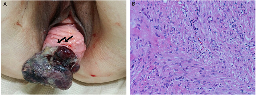
Fig. 1 (A) An irregular shaped tender protruding vaginal mass, black color with multiple focal necrosis, was found. The vagina mucosa was broken (arrows). (B) The high power photogram demonstrated the bundles of spindle cells interlacing in right angle. There was less than 2 mitotic figure (H&E, ×200).
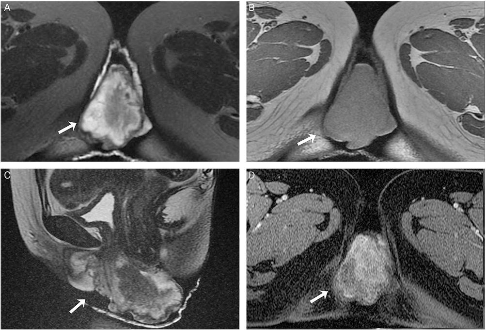
Fig. 2 Vaginal mass in a 43-year-old woman. (A) T2-weighted axial magnetic resonance (MR) image shows irregular shaped mass (arrow) in the vagina. The signal intensity of the mass is heterogenous and higher compared that of pelvic muscle. (B) T1-weighted axial MR image. The signal intensity of the mass (arrow) is similar to that of pelvic muscle. (C) The mass (arrow) has heterogenous high signal intensity on T2-weighted sagittal MR image. (D) The mass (arrow) has irregular enhancement on contrast enhanced T1-weighted MR image
Reference
-
1. Young SB, Rose PG, Reuter KL. Vaginal fibromyomata: two cases with preoperative assessment, resection, and reconstruction. Obstet Gynecol. 1991; 78:972–974.2. Freed SZ, Haleem SA, Wiener I, Feldman J. Bladder outlet obstruction caused by vaginal fibromyoma: the female prostate. J Urol. 1975; 113:30–31.3. Chakrabarti I, De A, Pati S. Vaginal leiomyoma. J Midlife Health. 2011; 2:42–43.4. Bennett HG, Ehrlich MM. Myoma of the vagina. Am J Obstet Gynecol. 1941; 42:314–320.5. Oruc S, Karaer O, Kurtul O. Coexistence of a prolapsed, pedunculated cervical myoma and pregnancy complications: a case report. J Reprod Med. 2004; 49:575–577.6. Liu MM. Fibromyoma of the vagina. Eur J Obstet Gynecol Reprod Biol. 1988; 29:321–328.7. Pavlica P, Bartolone A, Gaudiano C, Barozzi L. Female paraurethral leiomyoma: ultrasonographic and magnetic resonance imaging findings. Acta Radiol. 2004; 45:796–798.8. Shadbolt CL, Coakley FV, Qayyum A, Donat SM. MRI of vaginal leiomyomas. J Comput Assist Tomogr. 2001; 25:355–357.9. Ikeda R, Suga K, Suzuki K. MRI appearance of a leiomyoma of the female urethra. Clin Radiol. 2001; 56:76–79.10. Siegelman ES, Outwater EK. Tissue characterization in the female pelvis by means of MR imaging. Radiology. 1999; 212:5–18.11. Hubert KC, Remer EM, Rackley RR, Goldman HB. Clinical and magnetic resonance imaging characteristics of vaginal and paraurethral leiomyomas: can they be diagnosed before surgery? BJU Int. 2010; 105:1686–1688.12. Cobanoglu O, Gurkan Zorlu C, Ergun Y, Kutluay L. Leiomyosarcoma of the vagina. Eur J Obstet Gynecol Reprod Biol. 1996; 70:205–207.13. Haque AU, Moatasim A, Aslam F. Mitotically active leiomyoma: a word of caution. Int J Pathol. 2004; 2:38–41.14. Ahram J, Lemus R, Schiavello HJ. Leiomyosarcoma of the vagina: case report and literature review. Int J Gynecol Cancer. 2006; 16:884–891.15. Suh DH, Lim SY, Chung J, Choi HJ, Lee S, Park SY. Vaginal leiomyoma mimicking a malignant neoplasm on MR imaging. Korean J Obstet Gynecol. 2006; 49:2432–2437.
- Full Text Links
-
- Actions
-
Cited
- CITED
-
- Close
- Share
-
- Similar articles
-
- Vaginal leiomyoma mimicking a malignant neoplasm on MR imaging
- A Case of Suburethral Leiomyoma of the Vagina
- A Case of Leiomyoma of the Vaginal Wall in a Proximal Suburethral and Subtrigonal Location Causing Voiding Difficulty
- A Case of Leiomyoma of the Vagina
- A Case of Leiomyoma of the Uterus and Vagina
