Obstet Gynecol Sci.
2014 Jan;57(1):11-16.
Variety of prenatally diagnosed congenital heart disease in 22q11.2 deletion syndrome
- Affiliations
-
- 1Department of Obstetrics and Gynecology, University of Ulsan College of Medicine, Asan Medical Center, Seoul, Korea. hswon@amc.seoul.kr
Abstract
OBJECTIVE
To analyze the spectrum of prenatally diagnosed congenital heart disease in a Korean population with 22q11.2 deletion syndrome, and to provide guidelines for screening 22q11.2 deletion prenatally.
METHODS
This retrospective study evaluated 1,137 consecutive fetuses that had prenatal genetic testing for 22q11.2 deletion because of suspected congenital heart disease between September 2002 and December 2012, at Asan Medical Center, Seoul, Korea.
RESULTS
Main cardiovascular diseases in the 53 fetuses with confirmed 22q11.2 deletions were tetralogy of Fallot (n = 24, 45%), interrupted aortic arch (n = 10, 19%), ventricular septal defect (n = 5, 9%), double outlet right ventricle (n = 4, 8%), and coarctation of the aorta (n = 4, 8%). Other cardiac defects were rarely associated with 22q11.2 deletion. One fetus had persistent truncus arteriosus, one had aortic stenosis, and one had hypoplastic right heart syndrome. Two fetuses had normal intracardiac anatomy with an isolated right aortic arch, and one had an isolated bilateral superior vena cava.
CONCLUSION
A variety of congenital heart diseases were seen during the prenatal period. Conotruncal cardiac defects except transposition of great arteries were strongly associated with 22q11.2 deletion. When such anomalies are diagnosed by fetal echocardiography, genetic testing for 22q11.2 deletion should be offered. Even if less frequent deletion-related cardiac defects are detected, other related anomalies, such as thymic hypoplasia or aplasia, should be evaluated to rule out a 22q11.2 deletion.
MeSH Terms
-
Aorta, Thoracic
Aortic Coarctation
Aortic Valve Stenosis
Cardiovascular Diseases
Chungcheongnam-do
DiGeorge Syndrome*
Double Outlet Right Ventricle
Echocardiography
Fetus
Genetic Testing
Heart
Heart Defects, Congenital*
Heart Diseases
Heart Septal Defects, Ventricular
In Situ Hybridization, Fluorescence
Korea
Mass Screening
Retrospective Studies
Seoul
Tetralogy of Fallot
Transposition of Great Vessels
Truncus Arteriosus, Persistent
Vena Cava, Superior
Figure
-
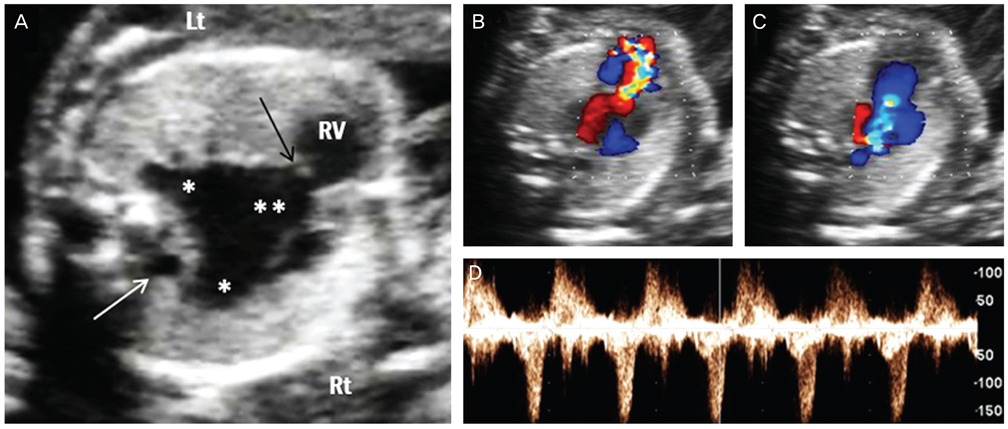
Fig. 1 Prenatal ultrasonographic findings of absent pulmonary valve syndrome. (A) Marked dilatation of main (**) and branch (*) pulmonary arteries with a rudimentary pulmonary valve (black arrow). Right aortic arch was also noted (white arrow). (B-D) Color and pulsed wave Doppler ultrasound showed a typical to-and-fro pattern of pulmonary stenosis and regurgitation. Lt, left; Rt, right; RV, right ventricle.
Reference
-
1. Wilson DI, Burn J, Scambler P, Goodship J. DiGeorge syndrome: part of CATCH 22. J Med Genet. 1993; 30:852–856.2. McElhinney DB, McDonald-McGinn D, Zackai EH, Goldmuntz E. Cardiovascular anomalies in patients diagnosed with a chromosome 22q11 deletion beyond 6 months of age. Pediatrics. 2001; 108:E104.3. Momma K. Cardiovascular anomalies associated with chromosome 22q11.2 deletion syndrome. Am J Cardiol. 2010; 105:1617–1624.4. Levy-Mozziconacci A, Wernert F, Scambler P, Rouault F, Metras D, Kreitman B, et al. Clinical and molecular study of DiGeorge sequence. Eur J Pediatr. 1994; 153:813–820.5. Ryan AK, Goodship JA, Wilson DI, Philip N, Levy A, Seidel H, et al. Spectrum of clinical features associated with interstitial chromosome 22q11 deletions: a European collaborative study. J Med Genet. 1997; 34:798–804.6. Matsuoka R, Kimura M, Scambler PJ, Morrow BE, Imamura S, Minoshima S, et al. Molecular and clinical study of 183 patients with conotruncal anomaly face syndrome. Hum Genet. 1998; 103:70–80.7. McDonald-McGinn DM, Kirschner R, Goldmuntz E, Sullivan K, Eicher P, Gerdes M, et al. The Philadelphia story: the 22q11.2 deletion: report on 250 patients. Genet Couns. 1999; 10:11–24.8. Oskarsdottir S, Persson C, Eriksson BO, Fasth A. Presenting phenotype in 100 children with the 22q11 deletion syndrome. Eur J Pediatr. 2005; 164:146–153.9. Park IS, Ko JK, Kim YH, Yoo HW, Seo EJ, Choi JY, et al. Cardiovascular anomalies in patients with chromosome 22q11.2 deletion: a Korean multicenter study. Int J Cardiol. 2007; 114:230–235.10. Levy-Mozziconacci A, Piquet C, Heurtevin PC, Philip N. Prenatal diagnosis of 22q11 microdeletion. Prenat Diagn. 1997; 17:1033–1037.11. Manji S, Roberson JR, Wiktor A, Vats S, Rush P, Diment S, et al. Prenatal diagnosis of 22q11.2 deletion when ultrasound examination reveals a heart defect. Genet Med. 2001; 3:65–66.12. Boudjemline Y, Fermont L, Le Bidois J, Lyonnet S, Sidi D, Bonnet D. Prevalence of 22q11 deletion in fetuses with conotruncal cardiac defects: a 6-year prospective study. J Pediatr. 2001; 138:520–524.13. Volpe P, Marasini M, Caruso G, Marzullo A, Buonadonna AL, Arciprete P, et al. 22q11 deletions in fetuses with malformations of the outflow tracts or interruption of the aortic arch: impact of additional ultrasound signs. Prenat Diagn. 2003; 23:752–757.14. Moore JW, Binder GA, Berry R. Prenatal diagnosis of aneuploidy and deletion 22q11.2 in fetuses with ultrasound detection of cardiac defects. Am J Obstet Gynecol. 2004; 191:2068–2073.15. Bretelle F, Beyer L, Pellissier MC, Missirian C, Sigaudy S, Gamerre M, et al. Prenatal and postnatal diagnosis of 22q11.2 deletion syndrome. Eur J Med Genet. 2010; 53:367–370.16. Momma K, Kondo C, Matsuoka R, Takao A. Cardiac anomalies associated with a chromosome 22q11 deletion in patients with conotruncal anomaly face syndrome. Am J Cardiol. 1996; 78:591–594.17. Lee MY, Won HS, Lee BS, Kim EA, Kim YH, Park JJ, et al. Prenatal diagnosis of common arterial trunk: a single-center's experience. Fetal Diagn Ther. 2013; 34:152–157.18. Momma K. Cardiovascular anomalies associated with chromosome 22q11.2 deletion. Int J Cardiol. 2007; 114:147–149.19. Driscoll DA. Prenatal diagnosis of the 22q11.2 deletion syndrome. Genet Med. 2001; 3:14–18.20. Zalel Y, Gamzu R, Mashiach S, Achiron R. The development of the fetal thymus: an in utero sonographic evaluation. Prenat Diagn. 2002; 22:114–117.21. Chaoui R, Kalache KD, Heling KS, Tennstedt C, Bommer C, Korner H. Absent or hypoplastic thymus on ultrasound: a marker for deletion 22q11.2 in fetal cardiac defects. Ultrasound Obstet Gynecol. 2002; 20:546–552.22. Li L, Bahtiyar MO, Buhimschi CS, Zou L, Zhou QC, Copel JA. Assessment of the fetal thymus by two- and three-dimensional ultrasound during normal human gestation and in fetuses with congenital heart defects. Ultrasound Obstet Gynecol. 2011; 37:404–409.23. Driscoll DA, Budarf ML, Emanuel BS. Antenatal diagnosis of DiGeorge syndrome. Lancet. 1991; 338:1390–1391.
- Full Text Links
-
- Actions
-
Cited
- CITED
-
- Close
- Share
-
- Similar articles
-
- Large Congenital Epigastric Hernia with DiGeorge Syndrome: A Case Report
- Genetic Syndromes associated with Congenital Heart Disease
- Prenatal screening of DiGeorge (22q11.2 deletion) syndrome by abnormalities of the great arteries among Thai pregnant women
- A Case of Bernard-Soulier Syndrome Associated with 22q11.2 Deletion Syndrome
- Prenatal Diagnosis of Tetralogy of Fallot Associated with Chromosome 22q11 Deletion
