Breast diseases during pregnancy and lactation
- Affiliations
-
- 1Department of Obstetrics and Gynecology, Miz Womens's Hospital, Daejeon, Korea.
- 2Department of Obstetrics and Gynecology, Bucheon St. Mary's Hospital, The Catholic University College of Medicine, Bucheon, Korea.
- 3Department of Obstetrics and Gynecology, Incheon Seoul Women's Hospital, Incheon, Korea.
- 4Department of Obstetrics and Gynecology, Chosun University College of Medicine, Gwangju, Korea. ogatg@chosun.ac.kr
Abstract
- Breast is a typical female sexual physiologic organ that is influenced by steroid hormone from menarche until menopause. Therefore various diseases can be developed by continuous action of estrogen and progesterone. Breast diseases are mainly categorized as benign and malignant. It is very important to distinguish the malignancy from breast diseases. However, it is very difficult to diagnose malignancy in pregnant and lactating women even though the same breast diseases took place. Therefore, we will review breast diseases such as breast carcinoma during pregnancy and lactation.
Keyword
MeSH Terms
Figure
-
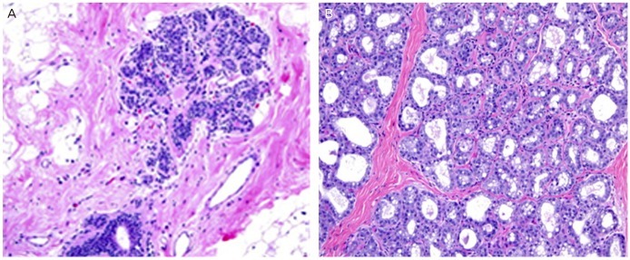
Fig. 1 Changes of breast tissue during lactation. (A) Terminal duct-lobular unit in non-pregnancy (H&E, ×200). (B) Dilated lobular acini with vacuoles and secretions can be seen during lactation (H&E, ×200).
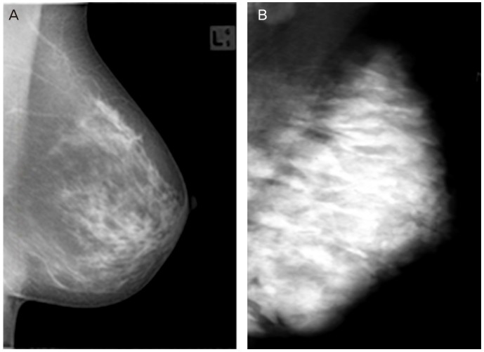
Fig. 2 Mammographic changes during lactation. (A) Type 2 American College of Radiology classification shows before pregnancy. (B) Mammogram during lactation shows a marked diffuse increase in density.
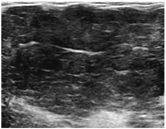
Fig. 3 Typical ultrasonographic feature during pregnancy shows diffuse enlargement of the non-fatty glandular component and global hypoechogenicity.
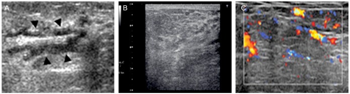
Fig. 4 Ultrasonographic changes during lactation. (A) Ultrasound (US) image shows irregular margined, hypoechoic dilated duct (black arrowhead). (B) US image reveals diffuse enlargement of the glandular component with diffuse hyperechogenicity. (C) Color Doppler US image reveals increased vascularity.
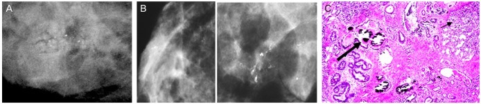
Fig. 5 Microcalcifications during lactation. (A) Image shows new cluster of indeterminate asymmetric microcalcification. (B) Craniocaudal spot-compression magnification mammograms: several clusters of heterogeneous and granular calcifications. Some clusters display linear distribution. (C) Photomicrograph of histopathologic specimen: a Coarse microcalcification group is seen in the dilated duct. The relatively bigger microcalcification (thick arrow) shows in the single duct with homogeneous and eosiophillic feature. The smaller microcalcification (thin arrow) is seen in the lobule (H&E, ×50).
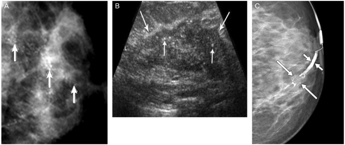
Fig. 6 A woman who presented with palpable mass and bloody nipple discharge at 24 weeks of pregnancy. (A) Magnification view mammogram of left breast in craniocaudal projection: extensive pleomorphic microcalcifications (arrows). (B) Corresponding longitudinal ultrasound image: irregular solid hypoechoic mass (long arrows) with internal calcifications (short arrows) corresponding to mammographic finding. (C) A mammogram of 30 weeks of pregnant woman presented with bloody nipple discharge: Multilobulated filling defect (long arrows), which focally expands duct. Proximal duct is dilated (short arrows).
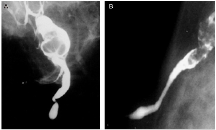
Fig. 7 (A) The galactogram in a lactating woman who presented with bloody nipple discharge: lobular filling defect is seen which is expected to be intraductal papilloma. (B) The galactograms in patients with papillomatosis showing intra-ductal growth in a separate duct.
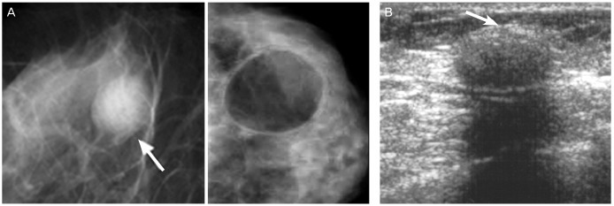
Fig. 8 Pseudolipoma type galactocele. (A) Mammography shows a 1.5 cm oval circumscribed mass (arrow) at the subareolar region. (B) Sonography shows a 1.5 cm oval circumscribed hypoechoic nodule with posterior shadowing. An echogenic rim (arrow) can be seen at the anterior margin.
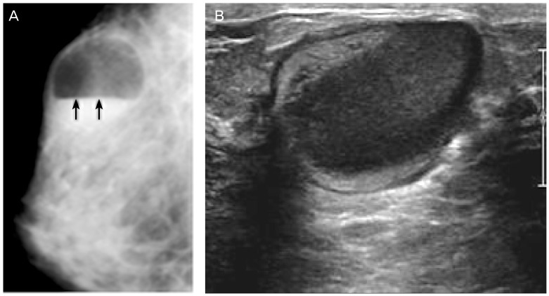
Fig. 9 Galactocele with fat-fluid level. (A) An oval circumscribed cystic mass (arrows) with fat-fluid level is imaged in mammogram. (B) Ultrasound image shows the fat-fluid level with high echogenicity of lipid component and low echogenicity of fluid in the same patient.
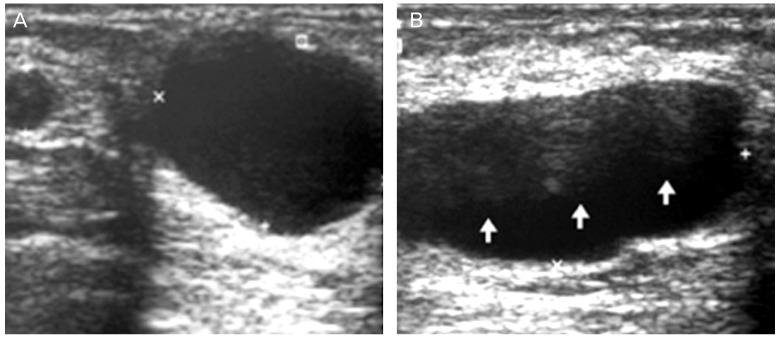
Fig. 10 Galactocele 6 months post delivery which shows various features. (A) Ultrasound (US) image reveals 2 cystic mass: typical galactocele with homogenous anechoic, acoustic attenuation and lateral edge shadowing in bigger cyst. (B) US image shows the lobulated, fat-fluid (arrows) galactocele.
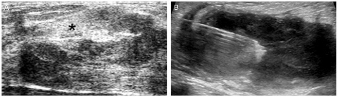
Fig. 11 Infected galactocele: lactating woman who presented with reddish skin changes in the breast. (A) Ultrasound image shows a heterogeneous echoic, irregular margined collection which was suspicious of abscess. (B) Fine-needle aspiration and culture was performed.
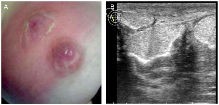
Fig. 12 Puerperal mastitis with abscess formation. (A) Lactational abscess grossly apparent secondary to flaming redness, hemorrhagic area, swelling, and peeling skin. (B) Ultrasound image shows large mass and purulent material was obtained by fine-needle aspiration.
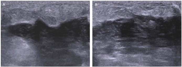
Fig. 13 Ultrasound findings in puerperal mastitis. Early stage mastitis shows various features that is presented with thickness of skin and subcutaneous layer, and irregular border between subcutaneous layer and parenchyme. (A) US shows irregular margin and hypoechoic lesion. (B) If abscess is develop, hypoechoic or anechoic fluid collections can be seen. Irregular margin and echoic lesion can be also seen along with acoustic enhancement.
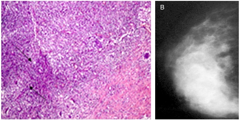
Fig. 14 Granulomatous mastitis after pregnancy. (A) Photomicrograph (enlarged, H&E, ×10) shows epitheloid and giant cell granulomas (arrows) in polymorphous inflammatory infiltrate. (B) Mammogram shows irregular enhanced mass almost filled the right breast.

Fig. 15 Lactating adenoma. (A) Ultrasound image demonstrates oval, well defined, regular margined mass. (B) Mammogram shows an oval circumscribed mass in the left lower breast. (C) The lobules are lined by actively secreting epithelial cells with vacuolated cytoplasm. Secretions may accumulate in the glands. The cells have basophilic cytoplasm, hyperchromatic nuclei with prominent nucleoli, and inconspicuous myoepithelial cell layer (H&E, ×400).
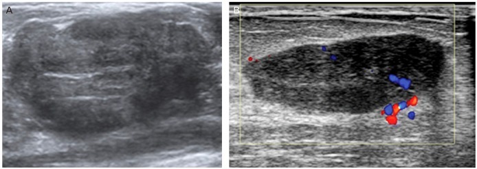
Fig. 16 Fibroadenoma. (A) Aside from cystic lesion, fibroadenoma shows internal echogenicity and fibroadenoma can not be distinguished from malignant lesion perfectly. (B) Color Doppler Ultrasound image shows a fibroadenoma with increased vascularity and lobulated hypoechoic mass.
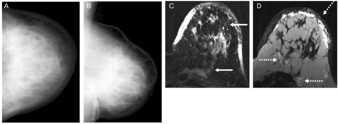
Fig. 17 Inflammatory carcinoma during pregnancy. (A) Craniocaudal view. (B) Mediolateral-oblique view: mammogram shows a mark diffuse increase in parenchymal density with skin thickening. (C) Subtraction 1 minute after bolus injection-the diffuse enhancement infiltrating the skin and the pectoralis muscle (continuous arrows). (D) T2-weighted image-edema in a cutaneous/subcutaneous, diffuse and prepectoral localization (discontinuous arrows).
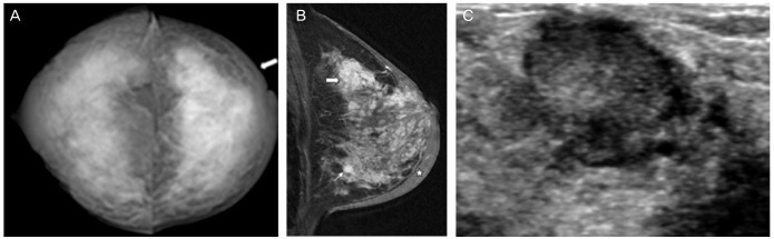
Fig. 18 Pregnancy-associated breast cancer in a lactating woman who is presented with paeau d'orange skin. (A) Bilateral craniocaudal mammogram: severe dense breast with hypertrophic skin. (B) Mediolateral-oblique view: multiple mass (thick and thin arrows). (C) Ultrasound image in a lactating woman presented with palpable mass 9 months post delivery: it shows taller than wide mixed echogeic lesion revealed with invasive ductal carcinoma.

Fig. 19 Atypical medullary carcinoma of the breast with cartilaginous metaplasia. (A) Ultrasound image reveals lobulated hypoechoic lesion in BRCA1 germline muation patient. (B) Higher magnification view of the previous slide shows the highly anaplastic tumor cells in a background of lymphoplasmacytic infiltrate (H&E, ×200).
Reference
-
1. Kopans DB. Breast imaging. 2th ed. Philadelphia (PA): Lippincott-Raven;1998.2. Salazar H, Tobon H, Josimovich JB. Developmental, gestational and postgestational modifications of the human breast. Clin Obstet Gynecol. 1975; 18:113–137. PMID: 1139807.
Article3. Rosen PP. Anatomic and physiologic morphology. In : Rosen PP, editor. Rosen's breast pathology. 2nd ed. Philadelphia (PA): Lippincott-Raven;2001. p. 1–21.4. Vorherr H. Human lactation and breast feeding. In : Larson BL, editor. Lactation: a comprehensive treatise. 2nd ed. New York (NY): Academic Press;1978. p. 182–280.6. Neville MC. Anatomy and physiology of lactation. Pediatr Clin North Am. 2001; 48:13–34. PMID: 11236721.
Article7. Hogge JP, De Paredes ES, Magnant CM, Lage J. Imaging and Management of Breast Masses During Pregnancy and Lactation. Breast J. 1999; 5:272–283. PMID: 11348301.
Article8. Liberman L, Giess CS, Dershaw DD, Deutch BM, Petrek JA. Imaging of pregnancy-associated breast cancer. Radiology. 1994; 191:245–248. PMID: 8134581.
Article9. Ahn BY, Kim HH, Moon WK, Pisano ED, Kim HS, Cha ES, et al. Pregnancy- and lactation-associated breast cancer: mammographic and sonographic findings. J Ultrasound Med. 2003; 22:491–497. PMID: 12751860.10. Samuels TH, Liu FF, Yaffe M, Haider M. Gestational breast cancer. Can Assoc Radiol J. 1998; 49:172–180. PMID: 9640283.11. Son EJ, Oh KK, Kim EK. Pregnancy-associated breast disease: radiologic features and diagnostic dilemmas. Yonsei Med J. 2006; 47:34–42. PMID: 16502483.
Article12. Yang WT, Dryden MJ, Gwyn K, Whitman GJ, Theriault R. Imaging of breast cancer diagnosed and treated with chemotherapy during pregnancy. Radiology. 2006; 239:52–60. PMID: 16484353.
Article13. Greskovich JF Jr, Macklis RM. Radiation therapy in pregnancy: risk calculation and risk minimization. Semin Oncol. 2000; 27:633–645. PMID: 11130470.14. Osei EK, Faulkner K. Fetal doses from radiological examinations. Br J Radiol. 1999; 72:773–780. PMID: 10624343.
Article15. Kopans DB. Mammography and radiation risk. In : Janower ML, Linton OW, editors. Radiation risk: a primer. Reston (VA): American College of Radiology;1996. p. 21–22.16. Kanal E, Borgstede JP, Barkovich AJ, Bell C, Bradley WG, Felmlee JP, et al. American College of Radiology White Paper on MR Safety. AJR Am J Roentgenol. 2002; 178:1335–1347. PMID: 12034593.
Article17. De Wilde JP, Rivers AW, Price DL. A review of the current use of magnetic resonance imaging in pregnancy and safety implications for the fetus. Prog Biophys Mol Biol. 2005; 87:335–353. PMID: 15556670.
Article18. Nagayama M, Watanabe Y, Okumura A, Amoh Y, Nakashita S, Dodo Y. Fast MR imaging in obstetrics. Radiographics. 2002; 22:563–580. PMID: 12006687.
Article19. Webb JA, Thomsen HS, Morcos SK. Members of Contrast Media Safety Committee of European Society of Urogenital Radiology (ESUR). The use of iodinated and gadolinium contrast media during pregnancy and lactation. Eur Radiol. 2005; 15:1234–1240. PMID: 15609057.
Article20. Mitre BK, Kanbour AI, Mauser N. Fine needle aspiration biopsy of breast carcinoma in pregnancy and lactation. Acta Cytol. 1997; 41:1121–1130. PMID: 9250309.
Article21. Gupta RK, McHutchison AG, Dowle CS, Simpson JS. Fine-needle aspiration cytodiagnosis of breast masses in pregnant and lactating women and its impact on management. Diagn Cytopathol. 1993; 9:156–159. PMID: 8513709.
Article22. Schackmuth EM, Harlow CL, Norton LW. Milk fistula: a complication after core breast biopsy. AJR Am J Roentgenol. 1993; 161:961–962. PMID: 8273635.
Article23. Stucker DT, Ikeda DM, Hartman AR, George TI, Nowels KW, Birdwell SL, et al. New bilateral microcalcifications at mammography in a postlactational woman: case report. Radiology. 2000; 217:247–250. PMID: 11012452.
Article24. Mercado CL, Koenigsberg TC, Hamele-Bena D, Smith SJ. Calcifications associated with lactational changes of the breast: mammographic findings with histologic correlation. AJR Am J Roentgenol. 2002; 179:685–689. PMID: 12185045.25. Giron GL, Boolbol SK, Gross J, Cohen JM, Feldman S. Postlactational microcalcifications. Breast J. 2004; 10:247–252. PMID: 15125754.
Article26. Lafreniere R. Bloody nipple discharge during pregnancy: a rationale for conservative treatment. J Surg Oncol. 1990; 43:228–230. PMID: 2325421.
Article27. Kline TS, Lash SR. The bleeding nipple of pregnancy and postpartum period: a cytologic and histologic study. Acta Cytol. 1964; 8:336–340. PMID: 14208372.28. O'Callaghan MA. Atypical discharge from the breast during pregnancy and/or lactation. Aust N Z J Obstet Gynaecol. 1981; 21:214–216. PMID: 6951562.29. Scott-Conner CEH. Diagnosing and managing breast disease during pregnancy and lactation. Medscape Womens Health. 1997; 2:1. PMID: 9746691.30. Rosen PP. Inflammatory and reactive tumors. In : Rosen PP, editor. Rosen's breast pathology. 2nd ed. Philadelphia (PA): Lippincott-Raven;2001. p. 29–63.31. Gómez A, Mata JM, Donoso L, Rams A. Galactocele: three distinctive radiographic appearances. Radiology. 1986; 158:43–44. PMID: 3940395.
Article32. Sawhney S, Petkovska L, Ramadan S, Al-Muhtaseb S, Jain R, Sheikh M. Sonographic appearances of galactoceles. J Clin Ultrasound. 2002; 30:18–22. PMID: 11807850.
Article33. Kim MJ, Kim EK, Park SY, Jung HK, Oh KK, Seok JY. Galactoceles mimicking suspicious solid masses on sonography. J Ultrasound Med. 2006; 25:145–151. PMID: 16439776.
Article34. Stevens K, Burrell HC, Evans AJ, Sibbering DM. The ultrasound appearances of galactocoeles. Br J Radiol. 1997; 70:239–241. PMID: 9166046.
Article35. Stafford I, Hernandez J, Laibl V, Sheffield J, Roberts S, Wendel G Jr. Community-acquired methicillin-resistant Staphylococcus aureus among patients with puerperal mastitis requiring hospitalization. Obstet Gynecol. 2008; 112:533–537. PMID: 18757649.
Article36. Marchant DJ. Inflammation of the breast. Obstet Gynecol Clin North Am. 2002; 29:89–102. PMID: 11892876.
Article37. Dixey JJ, Shanson DC, Williams TD, Rustin MH, Crook SJ, Midgley J, et al. Toxic-shock syndrome: four cases in a London hospital. Br Med J (Clin Res Ed). 1982; 285:342–343.
Article38. Karstrup S, Solvig J, Nolsoe CP, Nilsson P, Khattar S, Loren I, et al. Acute puerperal breast abscesses: US-guided drainage. Radiology. 1993; 188:807–809. PMID: 8351352.
Article39. Ulitzsch D, Nyman MK, Carlson RA. Breast abscess in lactating women: US-guided treatment. Radiology. 2004; 232:904–909. PMID: 15284435.
Article40. Eryilmaz R, Sahin M, Hakan Tekelioglu M, Daldal E. Management of lactational breast abscesses. Breast. 2005; 14:375–379. PMID: 16216739.
Article41. Memis A, Bilgen I, Ustun EE, Ozdemir N, Erhan Y, Kapkac M. Granulomatous mastitis: imaging findings with histopathologic correlation. Clin Radiol. 2002; 57:1001–1006. PMID: 12409111.
Article42. Yilmaz E, Lebe B, Usal C, Balci P. Mammographic and sonographic findings in the diagnosis of idiopathic granulomatous mastitis. Eur Radiol. 2001; 11:2236–2240. PMID: 11702165.
Article43. Han BK, Choe YH, Park JM, Moon WK, Ko YH, Yang JH, et al. Granulomatous mastitis: mammographic and sonographic appearances. AJR Am J Roentgenol. 1999; 173:317–320. PMID: 10430126.
Article44. Fletcher A, Magrath IM, Riddell RH, Talbot IC. Granulomatous mastitis: a report of seven cases. J Clin Pathol. 1982; 35:941–945. PMID: 6889612.
Article45. Asoglu O, Ozmen V, Karanlik H, Tunaci M, Cabioglu N, Igci A, et al. Feasibility of surgical management in patients with granulomatous mastitis. Breast J. 2005; 11:108–114. PMID: 15730456.
Article46. Tuncbilek N, Karakas HM, Okten OO. Imaging of granulomatous mastitis: assessment of three cases. Breast. 2004; 13:510–514. PMID: 15563860.
Article47. Lai EC, Chan WC, Ma TK, Tang AP, Poon CS, Leong HT. The role of conservative treatment in idiopathic granulomatous mastitis. Breast J. 2005; 11:454–456. PMID: 16297091.
Article48. Rosen PP. Breast tumors in children. In : Rosen PP, editor. Rosen's breast pathology. Philadelphia (PA): Lippincott-Raven;2001. p. 729–748.49. Dehner LP, Hill DA, Deschryver K. Pathology of the breast in children, adolescents, and young adults. Semin Diagn Pathol. 1999; 16:235–247. PMID: 10490200.50. Baker TP, Lenert JT, Parker J, Kemp B, Kushwaha A, Evans G, et al. Lactating adenoma: a diagnosis of exclusion. Breast J. 2001; 7:354–357. PMID: 11906446.
Article51. Saglam A, Can B. Coexistence of lactating adenoma and invasive ductal adenocarcinoma of the breast in a pregnant woman. J Clin Pathol. 2005; 58:87–89. PMID: 15623491.
Article52. Behrndt VS, Barbakoff D, Askin FB, Brem RF. Infarcted lactating adenoma presenting as a rapidly enlarging breast mass. AJR Am J Roentgenol. 1999; 173:933–935. PMID: 10511151.
Article53. Sumkin JH, Perrone AM, Harris KM, Nath ME, Amortegui AJ, Weinstein BJ. Lactating adenoma: US features and literature review. Radiology. 1998; 206:271–274. PMID: 9423682.
Article54. Yang WT, Suen M, Metreweli C. Lactating adenoma of the breast: antepartum and postpartum sonographic and color Doppler imaging appearances with histopathologic correlation. J Ultrasound Med. 1997; 16:145–147. PMID: 9166808.
Article55. Darling ML, Smith DN, Rhei E, Denison CM, Lester SC, Meyer JE. Lactating adenoma: sonographic features. Breast J. 2000; 6:252–256. PMID: 11348374.
Article56. Rosen PP. Fibroepithelial neoplasms. In : Rosen PP, editor. Rosen's breast pathology. 2nd ed. Philadelphia (PA): Lippincott-Raven;2001. p. 163–200.57. O'Hara MF, Page DL. Adenomas of the breast and ectopic breast under lactational influences. Hum Pathol. 1985; 16:707–712. PMID: 4007846.58. Novotny DB, Maygarden SJ, Shermer RW, Frable WJ. Fine needle aspiration of benign and malignant breast masses associated with pregnancy. Acta Cytol. 1991; 35:676–686. PMID: 1659095.59. Raju GC, Naraynsingh V. Infarction of fibroadenoma of the breast. J R Coll Surg Edinb. 1985; 30:162–163. PMID: 4045773.60. Majmudar B, Rosales-Quintana S. Infarction of breast fibroadenomas during pregnancy. JAMA. 1975; 231:963–964. PMID: 1173104.
Article61. Jimenez JF, Ryals RO, Cohen C. Spontaneous breast infarction associated with pregnancy presenting as a palpable mass. J Surg Oncol. 1986; 32:174–178. PMID: 3736054.
Article62. Petrek JA, Theriault RL. Pregnancy-associated breast cancer and subsequent pregnancy in breast cancer survivors. In : Harris JR, Lippman ME, Morrow M, Osborne CK, editors. Diseases of the breast. 3rd ed. Philadelphia (PA): Lippincott Williams & Williams;2004. p. 1035–1046.63. Ring AE, Smith IE, Ellis PA. Breast cancer and pregnancy. Ann Oncol. 2005; 16:1855–1860. PMID: 16030024.
Article64. Fuentes-Alburo A, Chavez-McGregor R, Ramirez Ugalde MT, De la Garza-Salazar JG. Early detection of breast cancer: who is responsible? 23rd Annual San Antonio Breast Cancer Symposium. Breast Cancer Res Treat. 2000; 64:abstract 205.65. Chiechi LM, Lobascio A, Loizzi P. Breast cancer in pregnancy. Resistance to screening. Minerva Ginecol. 1998; 50:301–304. PMID: 9808953.66. Nettleton J, Long J, Kuban D, Wu R, Shaefffer J, El-Mahdi A. Breast cancer during pregnancy: quantifying the risk of treatment delay. Obstet Gynecol. 1996; 87:414–418. PMID: 8598965.
Article67. Johannsson O, Loman N, Borg A, Olsson H. Pregnancy-associated breast cancer in BRCA1 and BRCA2 germline mutation carriers. Lancet. 1998; 352:1359–1360. PMID: 9802282.
Article68. Jernström H, Lerman C, Ghadirian P, Lynch HT, Weber B, Garber J, et al. Pregnancy and risk of early breast cancer in carriers of BRCA1 and BRCA2. Lancet. 1999; 354:1846–1850. PMID: 10584720.
Article69. Andrieu N, Goldgar DE, Easton DF, Rookus M, Brohet R, Antoniou AC, et al. Pregnancies, breast-feeding, and breast cancer risk in the International BRCA1/2 Carrier Cohort Study (IBCCS). J Natl Cancer Inst. 2006; 98:535–544. PMID: 16622123.
- Full Text Links
-
- Actions
-
Cited
- CITED
-
- Close
- Share
-
- Similar articles
-
- Breast lesions during pregnancy and lactation: a pictorial essay
- Breast cancer during pregnancy and lactation
- Ultrasonographic Findings of Breast Diseases During Pregnancy and Lactating Period
- Pregnancy and Lactation-associated Osteoporosis with Vertebral Compression Fracture
- Breast Cancer that was detected during Pregnancy
