Korean J Urol.
2009 Apr;50(4):413-416.
Castleman Disease Misdiagnosed as a Neoplasm of the Kidney
- Affiliations
-
- 1Department of Urology, Seoul Veterans Hospital, Seoul, Korea. urodoct@hotmail.com
- 2Department of Pathology, Seoul Veterans Hospital, Seoul, Korea.
Abstract
- Castleman disease, or angiofollicular lymph node hyperplasia, is a fairly rare benign tumor of lymphoid origin. Most cases tend to present as a mediastinal mass. We report a 58-year-old man with Castleman disease of the right perirenal space. This case was diagnosed preoperatively as nonconventional renal cell carcinoma (RCC) or renal oncocytoma because the enhancing mass abutted the renal cortex. The patient underwent a radical nephrectomy and a histopathological analysis showed the unicentric plasma cell type of Castleman disease. A preoperative diagnosis of Castleman disease is difficult; therefore, a surgical resection and a histological evaluation can provide an accurate diagnosis of this tumor. Taking this case into consideration, we suggest that Castleman disease should be included in the differential diagnosis of renal tumors.
Keyword
MeSH Terms
Figure
-
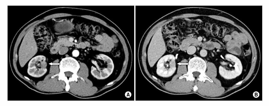
Fig. 1 CT scan showing a well-defined enhancing mass at the medial cortex of the right kidney (arrow). Arterial phase (A) and delayed phase (B).
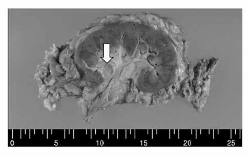
Fig. 2 Macroscopic view of the resected kidney. There is a 1.8×1.5×1.0 cm ovoid mass in the hilar soft tissue (arrow).
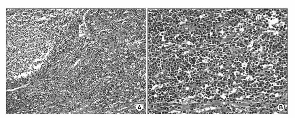
Fig. 3 Plasma cell type of Castleman disease. Microscopically, the interfollicular area contains a sheet of plasma cells [H&E, ×200 (A), ×400 (B)].
Reference
-
1. Castleman B, Iverson L, Menendez VP. Localized mediastinal lymphnode hyperplasia resembling thymoma. Cancer. 1956. 9:822–830.2. Hatano K, Fujita S, Tsujimoto Y, Takaya T, Honda M, Tsujimoto M, et al. Rare case of the hyaline vascular type of Castleman's disease of the kidney. Int J Urol. 2007. 14:1098–1100.3. Hsu HH, Chen YC, Fang JT, Huang CC, Shin LY, Ghuang CK. Unicentric Castleman's disease associated with hydronephrosis. Urology. 2000. 56:856.4. Waisberg J, Satake M, Yamagushi N, Matos LL, Waisberg DR, Artigiani Neto R, et al. Retroperitoneal unicentric Castleman's disease (giant lymph node hyperplasia): case report. Sao Paulo Med J. 2007. 125:253–255.5. Casper C. The aetiology and management of Castleman disease at 50 years: translating pathophysiology to patient care. Br J Haematol. 2005. 129:3–17.6. Kim TJ, Han JK, Kim YH, Kim TK, Choi BI. Castleman disease of the abdomen: imaging spectrum and clinicopathologic correlations. J Comput Assist Tomogr. 2001. 25:207–214.7. Rudloff U, Jacobson A, Morgenstern N, Chen Y, Lee BR. Castleman's disease of the urachus. Urology. 2004. 64:376–379.8. Lee JW, Park SY, Kim BS, Kim SJ, Son YW, Moon HS, et al. Retroperitoneal Castleman's disease incidentally detected due to urinary calculus. Korean J Urol. 2008. 49:186–189.9. Yamashita Y, Hirai T, Matsukawa T, Ogata I, Takahashi M. Radiological presentations of Castleman's disease. Comput Med Imaging Graph. 1993. 17:107–117.10. Kim YJ, Kim JH, Lee SJ, Kim JI. Hyaline-vascular variant of Castleman's disease in retroperitoneum. Korean J Urol. 2001. 42:668–670.
- Full Text Links
-
- Actions
-
Cited
- CITED
-
- Close
- Share
-
- Similar articles
-
- Castleman Disease in the Kidney and Retroperitoneum Mimicking Renal Cell Carcinoma with Retroperitoneal Lymphadenopathy: A Case Report
- A Case of Retroperitoneal Castleman`s Disease
- Hyaline-vascular Variant of Castleman's Disease in Retroperitoneum
- A case of Castleman disease that improved after kidney transplantation
- Development of Castleman Disease in the Paravertebral Space Mimicking a Neurogenic Tumor
