Kosin Med J.
2015 Jun;30(1):81-85. 10.7180/kmj.2015.30.1.81.
Tricuspid and pulmonary valve endocarditis associated with double-chambered right ventricle
- Affiliations
-
- 1Department of Cardiology, Eulji University Hospital, Eulji University School of Medicine, Daejeon, Korea. spyman2000@naver.com
- 2Department of Cardiovascular surgery, Eulji University Hospital, Eulji University School of Medicine, Daejeon, Korea.
- KMID: 2308541
- DOI: http://doi.org/10.7180/kmj.2015.30.1.81
Abstract
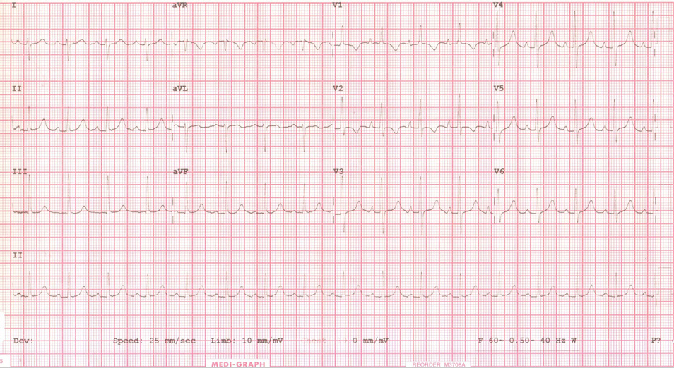
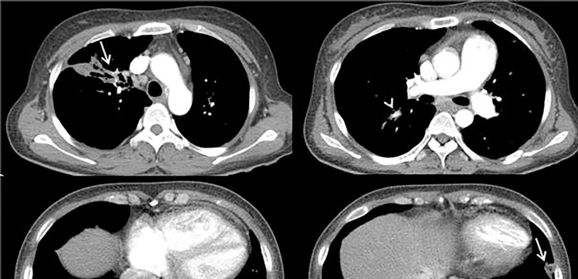
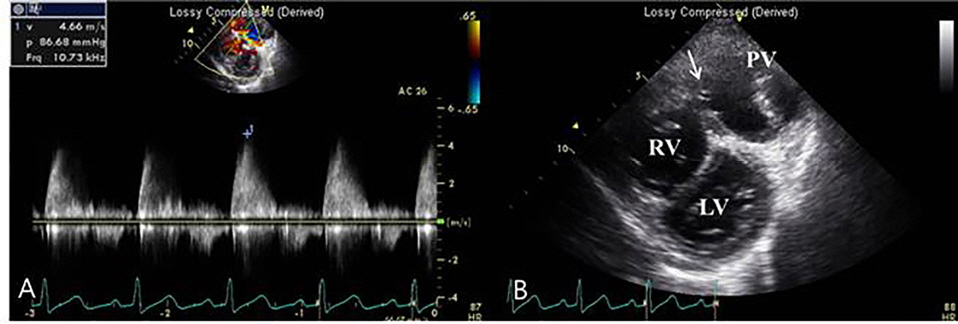
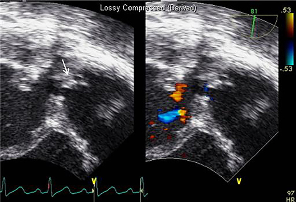
- We report a rare case of tricuspid valve and pulmonary valve endocarditis associated with a double-chambered right ventricle in an adult female with pulmonary artery aneurysm and septic pulmonary embolism by Streptococcus mitis. She was treated with aggressive antibiotic therapy followed by debridement of the infective lesion of tricuspid valve, pulmonary valve replacement using xenograft and resection of obstructing muscular bundles in right ventricle.
Keyword
MeSH Terms
Figure
Reference
-
References
1. Hoffman P, Wójcik AW, Rózan ′ski J, Siudalska H, Jakubowska E, Włodarska EK, et al. The role of echocardiography in diagnosing double chambered right ventricle in adults. Heart. 2004; 90:789–93.
Article2. López-Pardo F, Aguilera A, Villa M, Granado C, Campos A, Cisneros JM. Double-chambered right ventricle associated with mural and pulmonic valve endocarditis: description of a clinical case and review of the literature. Echocardiography. 2004; 21:171–3.
Article3. Telagh R, Alexi-Meskishvili V, Hetzer R, Lange PE, Berger F, Abdul-Khaliq H. Initial clinical manifestations and mid- and longterm results after surgical repair of double-chambered right ventricle in children and adults. Cardiol Young. 2008; 18:268–74.
Article4. Habib G, Hoen B, Tornos P, Thuny F, Prendergast B, Vilacosta I, et al. ESC Committee for Practice Guidelines.: Guidelines on the prevention, diagnosis, and treatment of infective endocarditis (new version 2009): the Task Force on the Prevention, Diagnosis, and Treatment of Infective Endocarditis of the European Society of Cardiology (ESC). Endorsed by the European Society of Clinical Microbiology and Infectious Diseases (ESCMID) and the International Society of Chemotherapy (ISC) for Infection and Cancer. Eur. Heart J. 2009; 30:2369–413.5. Choi YJ, Park SW. Characteristics of double-chambered right ventricle in adult patients. Korean J Intern Med. 2010; 25:147–53.
Article6. Simpson Jr WF, Sade RM, Crawford FA, Taylor AB, Fyfe DA. Double chambered right ventricle. Ann Thorac Surg. 1987; 44:7–10.7. Chaurasia AS, Nawale JM, Yemul MA. Double-Chambered Right Ventricle with Pulmonary Valve Endocarditis. Echocardiography. 2013; 30:167–70.
Article
- Full Text Links
- Actions
-
Cited
- CITED
-
- Close
- Share
- Similar articles
-
- Two-chambered right ventricle resulting from aberrant muscle bundles a case report
- A Case Report of Surgical Management of Tricuswpid Valve Endocarditis
- Pulmonary Thromboembolism and Infarction Caused by Right-Sided Infective Endocarditis in a Patient with Ventricular Septal Defect
- Tricuspid atresia: a re-evaluation and classification
- A Case of Double Chambered Right Ventricle with Congenital Right Ventricular True Diverticulum


