Kosin Med J.
2012 Dec;27(2):161-165. 10.7180/kmj.2012.27.2.161.
A Case of Spontaneous Bladder Rupture Mimicking Diabetic Nephropathy in a Patient with Type 2 Diabetes Mellitus
- Affiliations
-
- 1Department of Internal Medicine, College of Medicine, The Catholic University, Seoul, Korea. sangah@catholic.ac.kr
- KMID: 2308525
- DOI: http://doi.org/10.7180/kmj.2012.27.2.161
Abstract
- Spontaneous rupture of the urinary bladder is a rare clinical entity, with the incidence reported as 1 in 126,000 hospital admissions. It is often associated with malignancy, inflammatory lesions, irradiation, calculus, diverticulum, binge alcohol drinking, continuous bladder irrigation, and neurogenic bladder. In rare instances, bladder rupture occurs without obvious causes. This rare clinical condition is difficult to diagnose because of vague symptoms. High index of suspicion is needed as the mortality rate is high if untreated. A 37-year-old woman with uncontrolled type 2 diabetes, was admitted to the emergency room complaining of progressive abdominal distension and discomfort. She had a past history of tubo-ovarian and bladder abscess, and had undergone multiple surgical operations. From ascites fluid study, she was diagnosed as spontaneous bladder rupture. A transurethral catheter was inserted and the symptoms and signs resolved. Bladder rupture, mimicking acute kidney injury of diabetic nephropathy was disclosed without surgery.
MeSH Terms
Figure
-
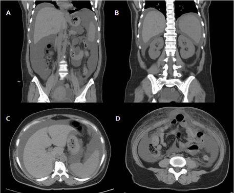
Fig. 1 (A-D) A abdominal computed tomography (CT) shows a large amount of ascites and focal ventral hernia of the small bowel loop, without hydronephrosis. The right and left kidney sizes are within normal limits, measuring 10.8 cm and 10.5 cm, respectively.
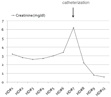
Fig. 2 Clinical course of the present case. The serum creatinine levels showed a dramatic decrease after transurethral catheter insertion.

Fig. 3 Follow up CT urography. (A) Ascites was resolved. (B) Focal ventral hernia of the small bowel loop is still noted. (C, D) Diverticulum-like outpouching was noted in the dome portion of the bladder without contrast leakage from the bladder.
Reference
-
1. Bastable JR, De Jode LR, Warren RP. Spontaneous rupture of the bladder. Br J Urol. 1959. 31:78–86.
Article2. Khan AU, Stern JM, Posalaky Z. Vesical necrosis associated with acute bladder overdistention. Urology. 1982. 19:197–199.
Article3. Horino T, Okazaki M, Nishikawa H, Takao T, Taniguchi Y, Morita T, et al. A case with spontaneous bladder rupture mimicking acute kidney injury. Clin Nephrol. 2009. 72:391–393.4. Manna R, Mirk P, Sallustio G, Brisinda G, Izzi D, La Regina M, et al. Hypercreatininemia and hyperglycemia: diabetic nephropathy or "inverted peritoneal auto-dialysis"? Clin Nephrol. 2005. 63:167–169.
Article5. Sullivan MJ, Lackner LH, Banowsky LH. Intraperitoneal extravasation of urine. BUN-serum creatinine disproportion. JAMA. 1972. 221:491–492.
Article6. Chan DP, Abujudeh HH, Cushing GL Jr, Novelline RA. CT cystography with multiplanar reformation for suspected bladder rupture: experience in 234 cases. AJR Am J Roentgenol. 2006. 187:1296–1302.
Article7. Mulkey AP Jr, Witherington R. Conservative management of vesical rupture. Urology. 1974. 4:426–430.
Article8. Mardani M, Shahzadi M, Rakhshani N, Rahnavardi M, Rezvani J, Sharifinejad A. Spontaneous perforation of urinary bladder secondary to Candida cystitis: acute abdomen of urologic origin. Surg Infect (Larchmt). 2008. 9:525–527.
Article9. Comiter CV, McDonald M, Minton J, Yalla SV. Fungal bezoar and bladder rupture secondary to candida tropicalis. Urology. 1996. 47:439–441.
Article10. Lee MH, Jung JY, Beak DH, Park YS, Yoo TK, Sung SA, et al. A case of sponataneous bladder rupture after a bout of heavy drinking. Korean J Med. 2009. 76:370–373.
- Full Text Links
-
- Actions
-
Cited
- CITED
-
- Close
- Share
-
- Similar articles
-
- Spontaneous Rupture of the Kidney as a Complication of Diabetes Mellitus
- Diabetic Nephropathy in Childhood and Adolescence (I) : Clinical Features
- Epidemiology and Clinical Course of Diabetic Nephropathy; Is There Any Differences in Prevalence and Incidence of Diabetic Nephropathy between Type 1 and Type 2 Diabetes Mellitus?
- Diet Therapy in Patients of Diabetic Nephropathy
- Glycemic Control in Diabetic Patients with Diabetic Nephropathy
